Magnesium »
PDB 3pip-3px4 »
3pmz »
Magnesium in PDB 3pmz: Crystal Structure of the Complex of Acetylcholine Binding Protein and D-Tubocurarine
Protein crystallography data
The structure of Crystal Structure of the Complex of Acetylcholine Binding Protein and D-Tubocurarine, PDB code: 3pmz
was solved by
T.T.Talley,
M.Harel,
J.G.Yamauchi,
Z.Radic,
S.Hansen,
T.Huxford,
P.W.Taylor,
with X-Ray Crystallography technique. A brief refinement statistics is given in the table below:
| Resolution Low / High (Å) | 114.71 / 2.44 |
| Space group | P 21 21 2 |
| Cell size a, b, c (Å), α, β, γ (°) | 142.737, 194.042, 101.615, 90.00, 90.00, 90.00 |
| R / Rfree (%) | 22.8 / 29.4 |
Magnesium Binding Sites:
Pages:
>>> Page 1 <<< Page 2, Binding sites: 11 - 16;Binding sites:
The binding sites of Magnesium atom in the Crystal Structure of the Complex of Acetylcholine Binding Protein and D-Tubocurarine (pdb code 3pmz). This binding sites where shown within 5.0 Angstroms radius around Magnesium atom.In total 16 binding sites of Magnesium where determined in the Crystal Structure of the Complex of Acetylcholine Binding Protein and D-Tubocurarine, PDB code: 3pmz:
Jump to Magnesium binding site number: 1; 2; 3; 4; 5; 6; 7; 8; 9; 10;
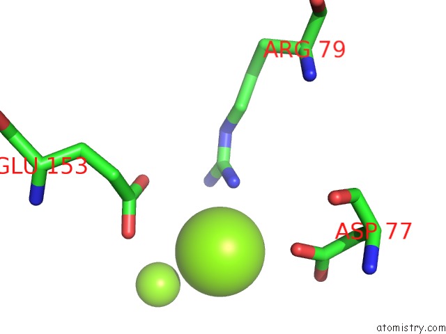
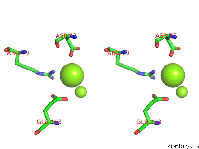
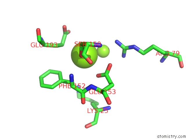
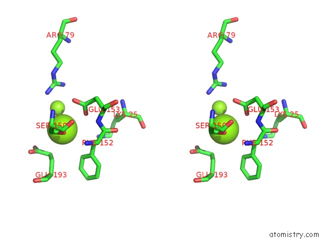
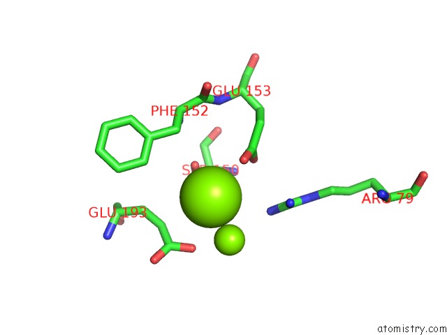
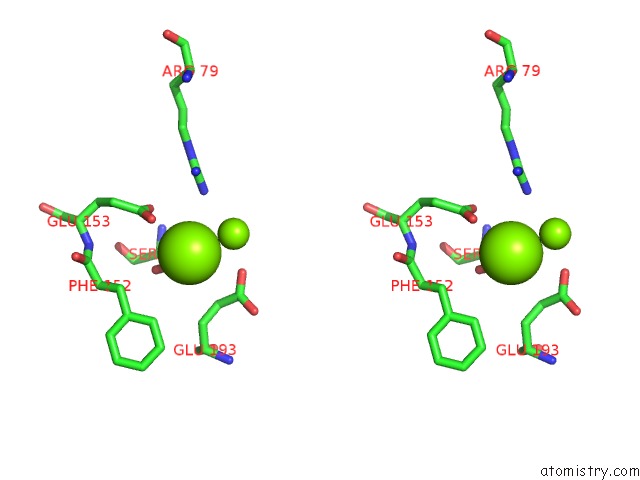
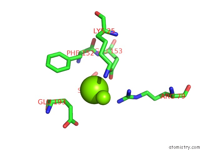
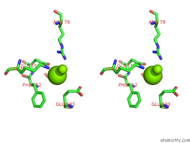
Magnesium binding site 1 out of 16 in 3pmz
Go back to
Magnesium binding site 1 out
of 16 in the Crystal Structure of the Complex of Acetylcholine Binding Protein and D-Tubocurarine
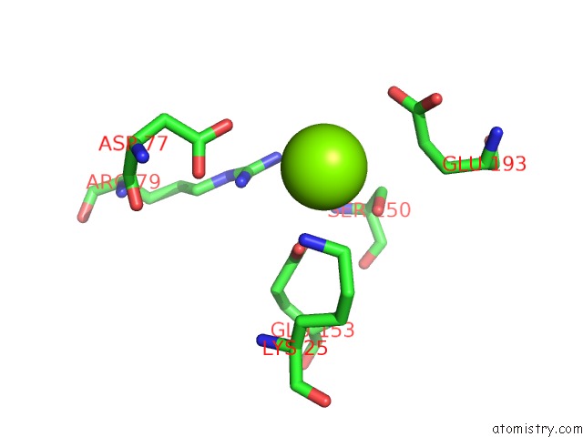
Mono view
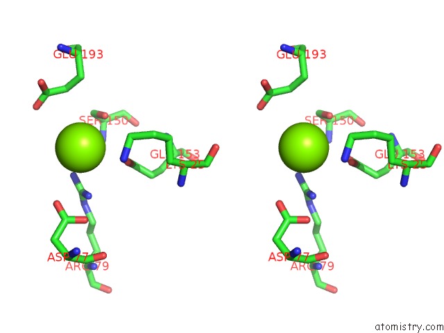
Stereo pair view
Mono view
Stereo pair view
A full contact list of Magnesium with other atoms in the Mg binding
site number 1 of Crystal Structure of the Complex of Acetylcholine Binding Protein and D-Tubocurarine within 5.0Å range:
|
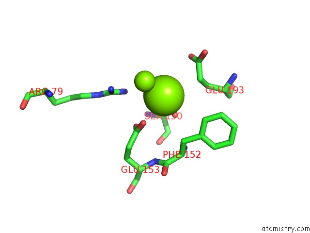
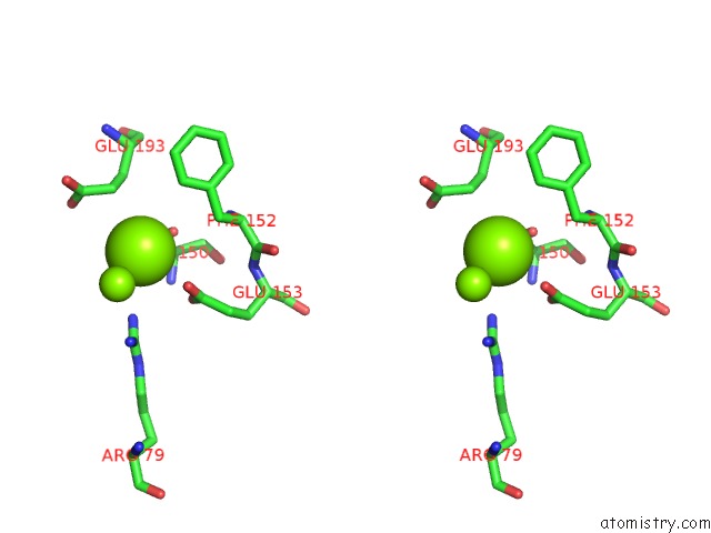
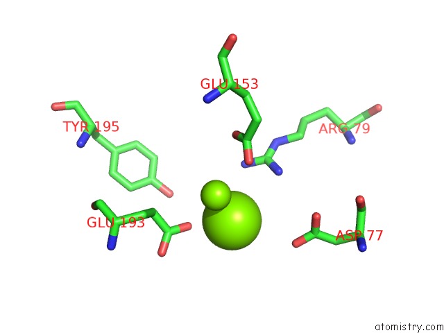
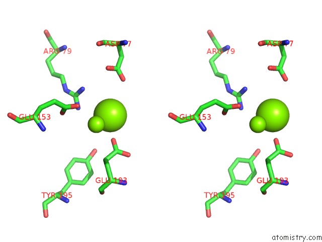
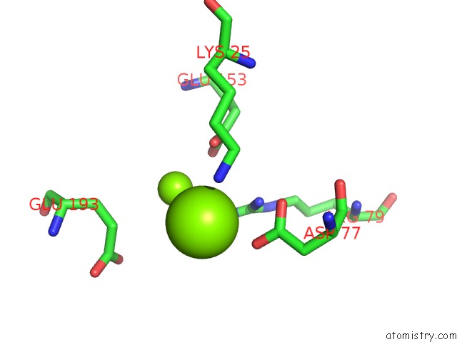
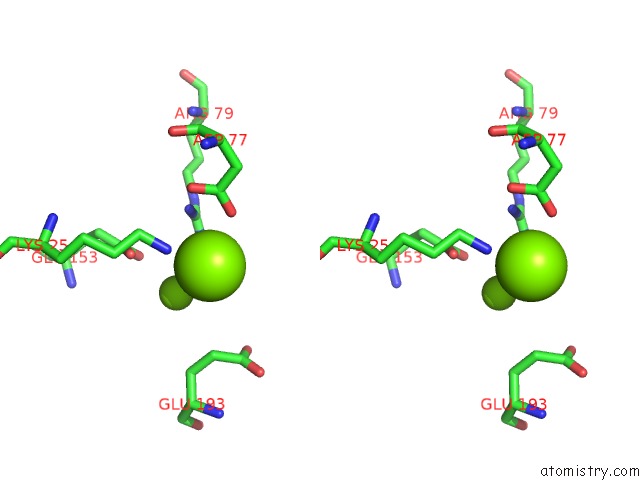
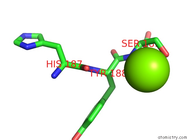
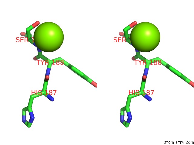
Magnesium binding site 2 out of 16 in 3pmz
Go back to
Magnesium binding site 2 out
of 16 in the Crystal Structure of the Complex of Acetylcholine Binding Protein and D-Tubocurarine
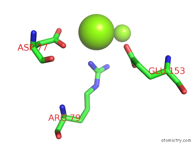
Mono view
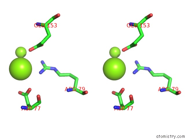
Stereo pair view
Mono view
Stereo pair view
A full contact list of Magnesium with other atoms in the Mg binding
site number 2 of Crystal Structure of the Complex of Acetylcholine Binding Protein and D-Tubocurarine within 5.0Å range:
|
Magnesium binding site 3 out of 16 in 3pmz
Go back to
Magnesium binding site 3 out
of 16 in the Crystal Structure of the Complex of Acetylcholine Binding Protein and D-Tubocurarine
Mono view
Stereo pair view
Mono view
Stereo pair view
A full contact list of Magnesium with other atoms in the Mg binding
site number 3 of Crystal Structure of the Complex of Acetylcholine Binding Protein and D-Tubocurarine within 5.0Å range:
|
Magnesium binding site 4 out of 16 in 3pmz
Go back to
Magnesium binding site 4 out
of 16 in the Crystal Structure of the Complex of Acetylcholine Binding Protein and D-Tubocurarine
Mono view
Stereo pair view
Mono view
Stereo pair view
A full contact list of Magnesium with other atoms in the Mg binding
site number 4 of Crystal Structure of the Complex of Acetylcholine Binding Protein and D-Tubocurarine within 5.0Å range:
|
Magnesium binding site 5 out of 16 in 3pmz
Go back to
Magnesium binding site 5 out
of 16 in the Crystal Structure of the Complex of Acetylcholine Binding Protein and D-Tubocurarine
Mono view
Stereo pair view
Mono view
Stereo pair view
A full contact list of Magnesium with other atoms in the Mg binding
site number 5 of Crystal Structure of the Complex of Acetylcholine Binding Protein and D-Tubocurarine within 5.0Å range:
|
Magnesium binding site 6 out of 16 in 3pmz
Go back to
Magnesium binding site 6 out
of 16 in the Crystal Structure of the Complex of Acetylcholine Binding Protein and D-Tubocurarine
Mono view
Stereo pair view
Mono view
Stereo pair view
A full contact list of Magnesium with other atoms in the Mg binding
site number 6 of Crystal Structure of the Complex of Acetylcholine Binding Protein and D-Tubocurarine within 5.0Å range:
|
Magnesium binding site 7 out of 16 in 3pmz
Go back to
Magnesium binding site 7 out
of 16 in the Crystal Structure of the Complex of Acetylcholine Binding Protein and D-Tubocurarine
Mono view
Stereo pair view
Mono view
Stereo pair view
A full contact list of Magnesium with other atoms in the Mg binding
site number 7 of Crystal Structure of the Complex of Acetylcholine Binding Protein and D-Tubocurarine within 5.0Å range:
|
Magnesium binding site 8 out of 16 in 3pmz
Go back to
Magnesium binding site 8 out
of 16 in the Crystal Structure of the Complex of Acetylcholine Binding Protein and D-Tubocurarine
Mono view
Stereo pair view
Mono view
Stereo pair view
A full contact list of Magnesium with other atoms in the Mg binding
site number 8 of Crystal Structure of the Complex of Acetylcholine Binding Protein and D-Tubocurarine within 5.0Å range:
|
Magnesium binding site 9 out of 16 in 3pmz
Go back to
Magnesium binding site 9 out
of 16 in the Crystal Structure of the Complex of Acetylcholine Binding Protein and D-Tubocurarine
Mono view
Stereo pair view
Mono view
Stereo pair view
A full contact list of Magnesium with other atoms in the Mg binding
site number 9 of Crystal Structure of the Complex of Acetylcholine Binding Protein and D-Tubocurarine within 5.0Å range:
|
Magnesium binding site 10 out of 16 in 3pmz
Go back to
Magnesium binding site 10 out
of 16 in the Crystal Structure of the Complex of Acetylcholine Binding Protein and D-Tubocurarine
Mono view
Stereo pair view
Mono view
Stereo pair view
A full contact list of Magnesium with other atoms in the Mg binding
site number 10 of Crystal Structure of the Complex of Acetylcholine Binding Protein and D-Tubocurarine within 5.0Å range:
|
Reference:
T.T.Talley,
M.Harel,
J.G.Yamauchi,
Z.Radic,
S.Hansen,
T.Huxford,
P.W.Taylor.
The Curare Alkaloids: Analyzing the Poses of Complexes with the Acetylcholine Binding Protein in Relation to Structure and Binding Energies To Be Published.
Page generated: Thu Aug 15 09:37:49 2024
Last articles
Fe in 2YXOFe in 2YRS
Fe in 2YXC
Fe in 2YNM
Fe in 2YVJ
Fe in 2YP1
Fe in 2YU2
Fe in 2YU1
Fe in 2YQB
Fe in 2YOO