Magnesium »
PDB 3pip-3px4 »
3pwg »
Magnesium in PDB 3pwg: Crystal Structure of the Mutant S29G.P34A of D-Glucarate Dehydratase From Escherichia Coli Complexed with Product 5-Keto-4-Deoxy-D- Glucarate
Protein crystallography data
The structure of Crystal Structure of the Mutant S29G.P34A of D-Glucarate Dehydratase From Escherichia Coli Complexed with Product 5-Keto-4-Deoxy-D- Glucarate, PDB code: 3pwg
was solved by
A.A.Fedorov,
E.V.Fedorov,
T.Lukk,
J.A.Gerlt,
S.C.Almo,
with X-Ray Crystallography technique. A brief refinement statistics is given in the table below:
| Resolution Low / High (Å) | 39.87 / 2.00 |
| Space group | P 21 21 21 |
| Cell size a, b, c (Å), α, β, γ (°) | 99.124, 130.658, 158.011, 90.00, 90.00, 90.00 |
| R / Rfree (%) | 23.1 / 28 |
Magnesium Binding Sites:
The binding sites of Magnesium atom in the Crystal Structure of the Mutant S29G.P34A of D-Glucarate Dehydratase From Escherichia Coli Complexed with Product 5-Keto-4-Deoxy-D- Glucarate
(pdb code 3pwg). This binding sites where shown within
5.0 Angstroms radius around Magnesium atom.
In total 4 binding sites of Magnesium where determined in the Crystal Structure of the Mutant S29G.P34A of D-Glucarate Dehydratase From Escherichia Coli Complexed with Product 5-Keto-4-Deoxy-D- Glucarate, PDB code: 3pwg:
Jump to Magnesium binding site number: 1; 2; 3; 4;
In total 4 binding sites of Magnesium where determined in the Crystal Structure of the Mutant S29G.P34A of D-Glucarate Dehydratase From Escherichia Coli Complexed with Product 5-Keto-4-Deoxy-D- Glucarate, PDB code: 3pwg:
Jump to Magnesium binding site number: 1; 2; 3; 4;
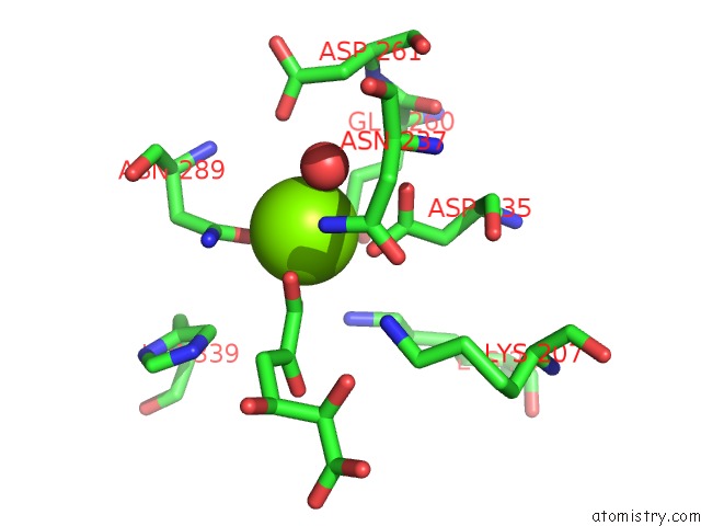
Magnesium binding site 1 out of 4 in 3pwg
Go back to
Magnesium binding site 1 out
of 4 in the Crystal Structure of the Mutant S29G.P34A of D-Glucarate Dehydratase From Escherichia Coli Complexed with Product 5-Keto-4-Deoxy-D- Glucarate

Mono view

Stereo pair view
Mono view
Stereo pair view
A full contact list of Magnesium with other atoms in the Mg binding
site number 1 of Crystal Structure of the Mutant S29G.P34A of D-Glucarate Dehydratase From Escherichia Coli Complexed with Product 5-Keto-4-Deoxy-D- Glucarate within 5.0Å range:
|
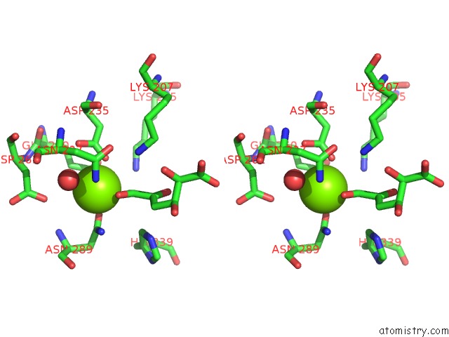
Magnesium binding site 2 out of 4 in 3pwg
Go back to
Magnesium binding site 2 out
of 4 in the Crystal Structure of the Mutant S29G.P34A of D-Glucarate Dehydratase From Escherichia Coli Complexed with Product 5-Keto-4-Deoxy-D- Glucarate
Mono view
Stereo pair view
Mono view
Stereo pair view
A full contact list of Magnesium with other atoms in the Mg binding
site number 2 of Crystal Structure of the Mutant S29G.P34A of D-Glucarate Dehydratase From Escherichia Coli Complexed with Product 5-Keto-4-Deoxy-D- Glucarate within 5.0Å range:
|
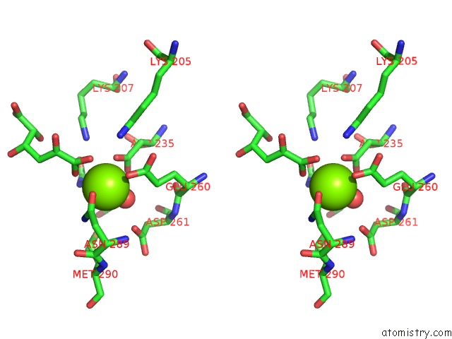
Magnesium binding site 3 out of 4 in 3pwg
Go back to
Magnesium binding site 3 out
of 4 in the Crystal Structure of the Mutant S29G.P34A of D-Glucarate Dehydratase From Escherichia Coli Complexed with Product 5-Keto-4-Deoxy-D- Glucarate

Mono view
Stereo pair view
Mono view
Stereo pair view
A full contact list of Magnesium with other atoms in the Mg binding
site number 3 of Crystal Structure of the Mutant S29G.P34A of D-Glucarate Dehydratase From Escherichia Coli Complexed with Product 5-Keto-4-Deoxy-D- Glucarate within 5.0Å range:
|
Magnesium binding site 4 out of 4 in 3pwg
Go back to
Magnesium binding site 4 out
of 4 in the Crystal Structure of the Mutant S29G.P34A of D-Glucarate Dehydratase From Escherichia Coli Complexed with Product 5-Keto-4-Deoxy-D- Glucarate

Mono view

Stereo pair view
Mono view
Stereo pair view
A full contact list of Magnesium with other atoms in the Mg binding
site number 4 of Crystal Structure of the Mutant S29G.P34A of D-Glucarate Dehydratase From Escherichia Coli Complexed with Product 5-Keto-4-Deoxy-D- Glucarate within 5.0Å range:
|
Reference:
A.A.Fedorov,
E.V.Fedorov,
T.Lukk,
J.A.Gerlt,
S.C.Almo.
Crystal Structure of the Mutant S29G.P34A of D-Glucarate Dehydratase From Escherichia Coli Complexed with Product 5-Keto-4-Deoxy-D-Glucarate To Be Published.
Page generated: Thu Aug 15 09:45:19 2024
Last articles
F in 7JL0F in 7JH2
F in 7JK8
F in 7JHW
F in 7JHD
F in 7I18
F in 7I2F
F in 7I2M
F in 7I2A
F in 7I2D