Magnesium »
PDB 3py8-3q86 »
3q3x »
Magnesium in PDB 3q3x: Crystal Structure of the Main Protease (3C) From Human Enterovirus B EV93
Enzymatic activity of Crystal Structure of the Main Protease (3C) From Human Enterovirus B EV93
All present enzymatic activity of Crystal Structure of the Main Protease (3C) From Human Enterovirus B EV93:
3.4.22.28;
3.4.22.28;
Protein crystallography data
The structure of Crystal Structure of the Main Protease (3C) From Human Enterovirus B EV93, PDB code: 3q3x
was solved by
L.Costenaro,
M.Sola,
B.Coutard,
H.Norder,
B.Canard,
M.Coll,
with X-Ray Crystallography technique. A brief refinement statistics is given in the table below:
| Resolution Low / High (Å) | 46.52 / 1.90 |
| Space group | P 1 21 1 |
| Cell size a, b, c (Å), α, β, γ (°) | 39.072, 65.216, 66.355, 90.00, 90.67, 90.00 |
| R / Rfree (%) | 14.8 / 21 |
Magnesium Binding Sites:
The binding sites of Magnesium atom in the Crystal Structure of the Main Protease (3C) From Human Enterovirus B EV93
(pdb code 3q3x). This binding sites where shown within
5.0 Angstroms radius around Magnesium atom.
In total 2 binding sites of Magnesium where determined in the Crystal Structure of the Main Protease (3C) From Human Enterovirus B EV93, PDB code: 3q3x:
Jump to Magnesium binding site number: 1; 2;
In total 2 binding sites of Magnesium where determined in the Crystal Structure of the Main Protease (3C) From Human Enterovirus B EV93, PDB code: 3q3x:
Jump to Magnesium binding site number: 1; 2;
Magnesium binding site 1 out of 2 in 3q3x
Go back to
Magnesium binding site 1 out
of 2 in the Crystal Structure of the Main Protease (3C) From Human Enterovirus B EV93
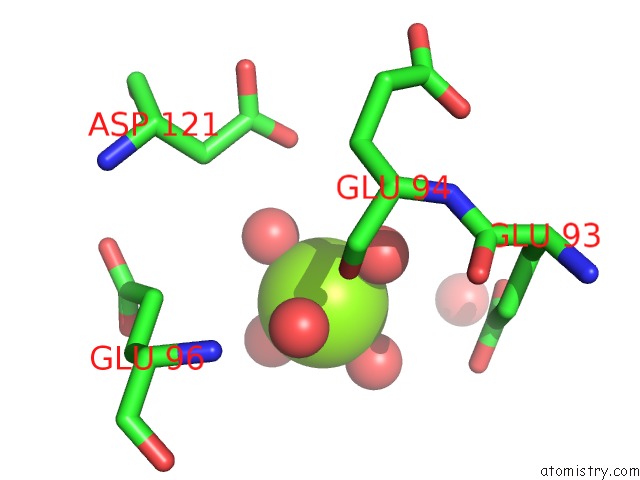
Mono view
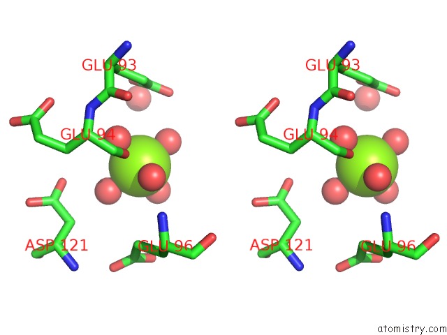
Stereo pair view
Mono view
Stereo pair view
A full contact list of Magnesium with other atoms in the Mg binding
site number 1 of Crystal Structure of the Main Protease (3C) From Human Enterovirus B EV93 within 5.0Å range:
|
Magnesium binding site 2 out of 2 in 3q3x
Go back to
Magnesium binding site 2 out
of 2 in the Crystal Structure of the Main Protease (3C) From Human Enterovirus B EV93
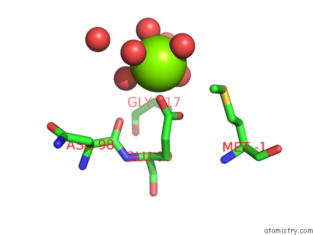
Mono view
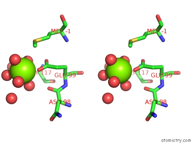
Stereo pair view
Mono view
Stereo pair view
A full contact list of Magnesium with other atoms in the Mg binding
site number 2 of Crystal Structure of the Main Protease (3C) From Human Enterovirus B EV93 within 5.0Å range:
|
Reference:
L.Costenaro,
Z.Kaczmarska,
C.Arnan,
R.Janowski,
B.Coutard,
M.Sola,
A.E.Gorbalenya,
H.Norder,
B.Canard,
M.Coll.
Structural Basis For Antiviral Inhibition of the Main Protease, 3C, From Human Enterovirus 93. J.Virol. V. 85 10764 2011.
ISSN: ISSN 0022-538X
PubMed: 21835784
DOI: 10.1128/JVI.05062-11
Page generated: Thu Aug 15 09:53:51 2024
ISSN: ISSN 0022-538X
PubMed: 21835784
DOI: 10.1128/JVI.05062-11
Last articles
Cl in 3SH3Cl in 3SGW
Cl in 3SGL
Cl in 3SG8
Cl in 3SG9
Cl in 3SFX
Cl in 3SFD
Cl in 3SFH
Cl in 3SFP
Cl in 3SFF