Magnesium »
PDB 3py8-3q86 »
3q6j »
Magnesium in PDB 3q6j: Structural Basis For Carbon Dioxide Binding By 2-Ketopropyl Coenzyme M Oxidoreductase/Carboxylase
Enzymatic activity of Structural Basis For Carbon Dioxide Binding By 2-Ketopropyl Coenzyme M Oxidoreductase/Carboxylase
All present enzymatic activity of Structural Basis For Carbon Dioxide Binding By 2-Ketopropyl Coenzyme M Oxidoreductase/Carboxylase:
1.8.1.5;
1.8.1.5;
Protein crystallography data
The structure of Structural Basis For Carbon Dioxide Binding By 2-Ketopropyl Coenzyme M Oxidoreductase/Carboxylase, PDB code: 3q6j
was solved by
A.S.Pandey,
D.W.Mulder,
S.A.Ensign,
J.W.Peters,
with X-Ray Crystallography technique. A brief refinement statistics is given in the table below:
| Resolution Low / High (Å) | 8.00 / 1.92 |
| Space group | P 1 21 1 |
| Cell size a, b, c (Å), α, β, γ (°) | 88.466, 59.803, 105.463, 90.00, 102.28, 90.00 |
| R / Rfree (%) | 16.4 / 22.1 |
Magnesium Binding Sites:
The binding sites of Magnesium atom in the Structural Basis For Carbon Dioxide Binding By 2-Ketopropyl Coenzyme M Oxidoreductase/Carboxylase
(pdb code 3q6j). This binding sites where shown within
5.0 Angstroms radius around Magnesium atom.
In total 2 binding sites of Magnesium where determined in the Structural Basis For Carbon Dioxide Binding By 2-Ketopropyl Coenzyme M Oxidoreductase/Carboxylase, PDB code: 3q6j:
Jump to Magnesium binding site number: 1; 2;
In total 2 binding sites of Magnesium where determined in the Structural Basis For Carbon Dioxide Binding By 2-Ketopropyl Coenzyme M Oxidoreductase/Carboxylase, PDB code: 3q6j:
Jump to Magnesium binding site number: 1; 2;
Magnesium binding site 1 out of 2 in 3q6j
Go back to
Magnesium binding site 1 out
of 2 in the Structural Basis For Carbon Dioxide Binding By 2-Ketopropyl Coenzyme M Oxidoreductase/Carboxylase
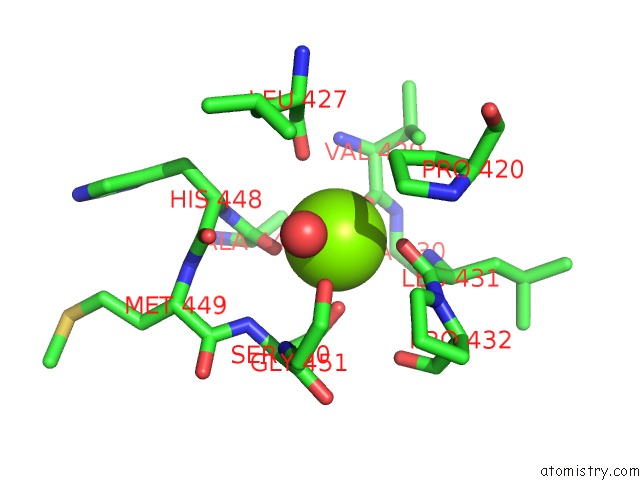
Mono view
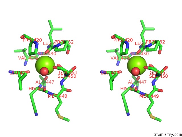
Stereo pair view
Mono view
Stereo pair view
A full contact list of Magnesium with other atoms in the Mg binding
site number 1 of Structural Basis For Carbon Dioxide Binding By 2-Ketopropyl Coenzyme M Oxidoreductase/Carboxylase within 5.0Å range:
|
Magnesium binding site 2 out of 2 in 3q6j
Go back to
Magnesium binding site 2 out
of 2 in the Structural Basis For Carbon Dioxide Binding By 2-Ketopropyl Coenzyme M Oxidoreductase/Carboxylase
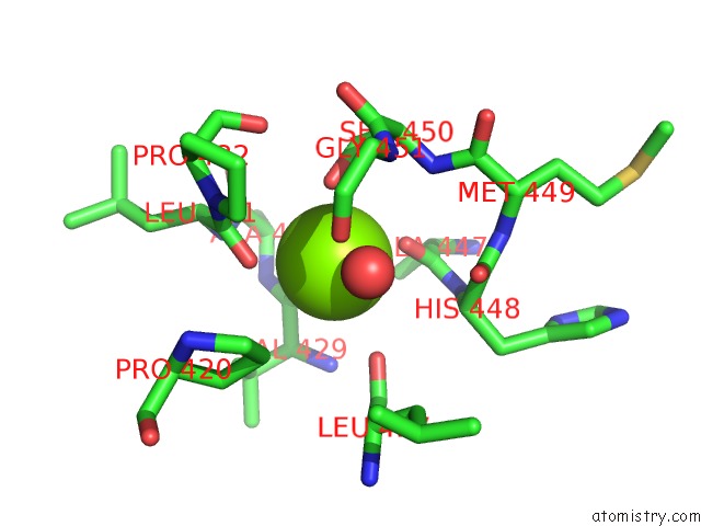
Mono view
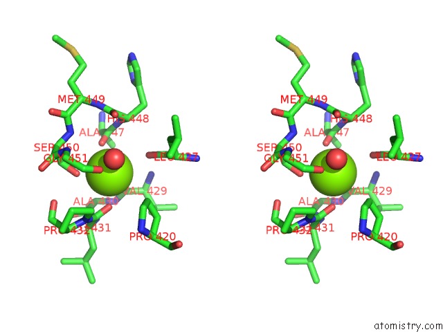
Stereo pair view
Mono view
Stereo pair view
A full contact list of Magnesium with other atoms in the Mg binding
site number 2 of Structural Basis For Carbon Dioxide Binding By 2-Ketopropyl Coenzyme M Oxidoreductase/Carboxylase within 5.0Å range:
|
Reference:
A.S.Pandey,
D.W.Mulder,
S.A.Ensign,
J.W.Peters.
Structural Basis For Carbon Dioxide Binding By 2-Ketopropyl Coenzyme M Oxidoreductase/Carboxylase. Febs Lett. V. 585 459 2011.
ISSN: ISSN 0014-5793
PubMed: 21192936
DOI: 10.1016/J.FEBSLET.2010.12.035
Page generated: Thu Aug 15 09:57:36 2024
ISSN: ISSN 0014-5793
PubMed: 21192936
DOI: 10.1016/J.FEBSLET.2010.12.035
Last articles
Cl in 8A4TCl in 8A51
Cl in 8A29
Cl in 8A4Q
Cl in 8A4G
Cl in 8A1Z
Cl in 8A1O
Cl in 8A21
Cl in 8A1M
Cl in 8A1Q