Magnesium »
PDB 3py8-3q86 »
3q7e »
Magnesium in PDB 3q7e: Crystal Structure of Rat Protein Arginine Methyltransferase 1 (PRMT1) M48L Mutant
Protein crystallography data
The structure of Crystal Structure of Rat Protein Arginine Methyltransferase 1 (PRMT1) M48L Mutant, PDB code: 3q7e
was solved by
S.J.Johnson,
P.J.Porter,
J.M.Hevel,
with X-Ray Crystallography technique. A brief refinement statistics is given in the table below:
| Resolution Low / High (Å) | 27.69 / 2.20 |
| Space group | P 41 2 2 |
| Cell size a, b, c (Å), α, β, γ (°) | 87.307, 87.307, 143.290, 90.00, 90.00, 90.00 |
| R / Rfree (%) | 20.1 / 24.5 |
Magnesium Binding Sites:
The binding sites of Magnesium atom in the Crystal Structure of Rat Protein Arginine Methyltransferase 1 (PRMT1) M48L Mutant
(pdb code 3q7e). This binding sites where shown within
5.0 Angstroms radius around Magnesium atom.
In total only one binding site of Magnesium was determined in the Crystal Structure of Rat Protein Arginine Methyltransferase 1 (PRMT1) M48L Mutant, PDB code: 3q7e:
In total only one binding site of Magnesium was determined in the Crystal Structure of Rat Protein Arginine Methyltransferase 1 (PRMT1) M48L Mutant, PDB code: 3q7e:
Magnesium binding site 1 out of 1 in 3q7e
Go back to
Magnesium binding site 1 out
of 1 in the Crystal Structure of Rat Protein Arginine Methyltransferase 1 (PRMT1) M48L Mutant
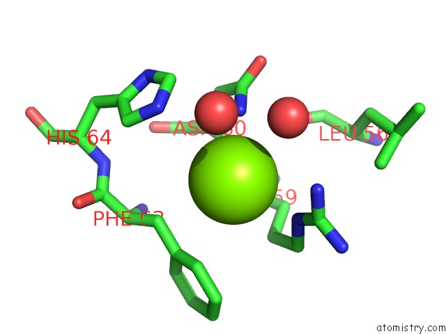
Mono view
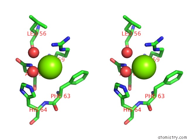
Stereo pair view
Mono view
Stereo pair view
A full contact list of Magnesium with other atoms in the Mg binding
site number 1 of Crystal Structure of Rat Protein Arginine Methyltransferase 1 (PRMT1) M48L Mutant within 5.0Å range:
|
Reference:
S.Gui,
W.L.Wooderchak,
M.P.Daly,
P.J.Porter,
S.J.Johnson,
J.M.Hevel.
Investigation of the Molecular Origins of Protein-Arginine Methyltransferase I (PRMT1) Product Specificity Reveals A Role For Two Conserved Methionine Residues. J.Biol.Chem. V. 286 29118 2011.
ISSN: ISSN 0021-9258
PubMed: 21697082
DOI: 10.1074/JBC.M111.224097
Page generated: Thu Aug 15 09:58:12 2024
ISSN: ISSN 0021-9258
PubMed: 21697082
DOI: 10.1074/JBC.M111.224097
Last articles
Zn in 9MJ5Zn in 9HNW
Zn in 9G0L
Zn in 9FNE
Zn in 9DZN
Zn in 9E0I
Zn in 9D32
Zn in 9DAK
Zn in 8ZXC
Zn in 8ZUF