Magnesium »
PDB 3q89-3qpo »
3qof »
Magnesium in PDB 3qof: Crystal Structure of the Cytosolic Domain of Human Atlastin-1 in Complex with Gdp, Orthorhombic Form
Protein crystallography data
The structure of Crystal Structure of the Cytosolic Domain of Human Atlastin-1 in Complex with Gdp, Orthorhombic Form, PDB code: 3qof
was solved by
X.Liu,
with X-Ray Crystallography technique. A brief refinement statistics is given in the table below:
| Resolution Low / High (Å) | 40.00 / 2.80 |
| Space group | P 21 21 21 |
| Cell size a, b, c (Å), α, β, γ (°) | 104.742, 133.445, 176.266, 90.00, 90.00, 90.00 |
| R / Rfree (%) | 22.5 / 26.7 |
Magnesium Binding Sites:
The binding sites of Magnesium atom in the Crystal Structure of the Cytosolic Domain of Human Atlastin-1 in Complex with Gdp, Orthorhombic Form
(pdb code 3qof). This binding sites where shown within
5.0 Angstroms radius around Magnesium atom.
In total 4 binding sites of Magnesium where determined in the Crystal Structure of the Cytosolic Domain of Human Atlastin-1 in Complex with Gdp, Orthorhombic Form, PDB code: 3qof:
Jump to Magnesium binding site number: 1; 2; 3; 4;
In total 4 binding sites of Magnesium where determined in the Crystal Structure of the Cytosolic Domain of Human Atlastin-1 in Complex with Gdp, Orthorhombic Form, PDB code: 3qof:
Jump to Magnesium binding site number: 1; 2; 3; 4;
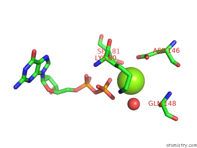
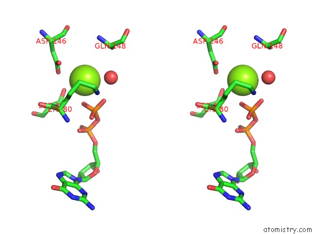
Magnesium binding site 1 out of 4 in 3qof
Go back to
Magnesium binding site 1 out
of 4 in the Crystal Structure of the Cytosolic Domain of Human Atlastin-1 in Complex with Gdp, Orthorhombic Form
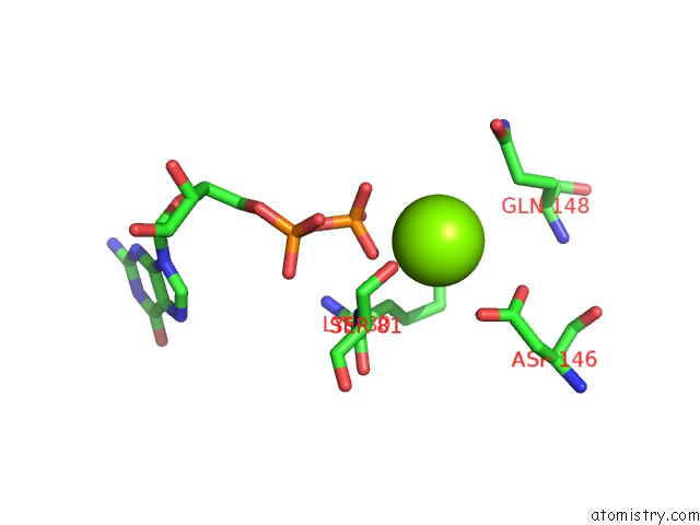
Mono view
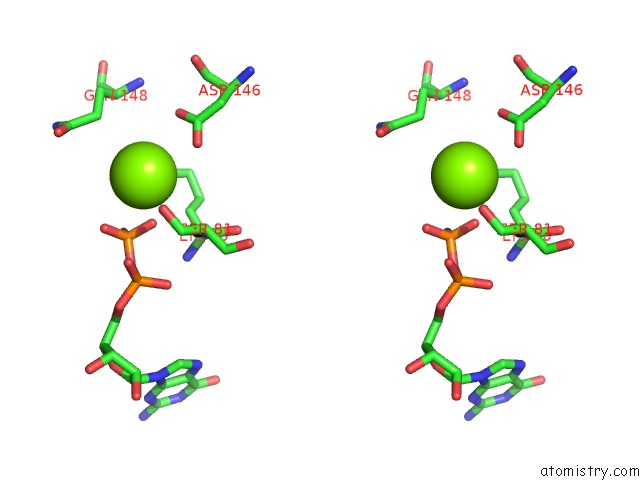
Stereo pair view
Mono view
Stereo pair view
A full contact list of Magnesium with other atoms in the Mg binding
site number 1 of Crystal Structure of the Cytosolic Domain of Human Atlastin-1 in Complex with Gdp, Orthorhombic Form within 5.0Å range:
|
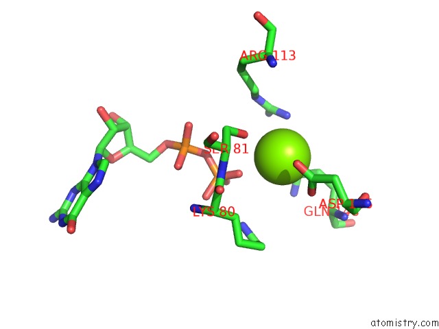
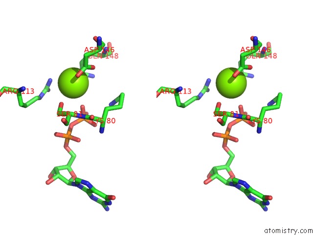
Magnesium binding site 2 out of 4 in 3qof
Go back to
Magnesium binding site 2 out
of 4 in the Crystal Structure of the Cytosolic Domain of Human Atlastin-1 in Complex with Gdp, Orthorhombic Form
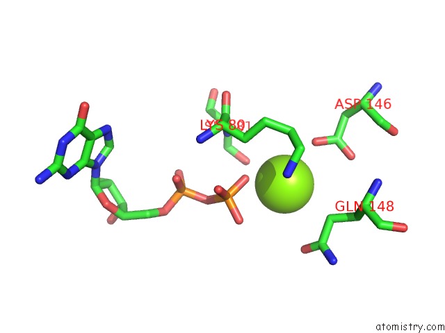
Mono view
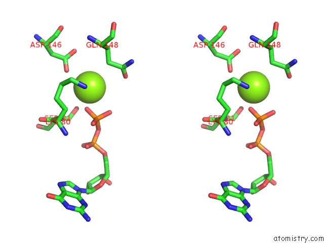
Stereo pair view
Mono view
Stereo pair view
A full contact list of Magnesium with other atoms in the Mg binding
site number 2 of Crystal Structure of the Cytosolic Domain of Human Atlastin-1 in Complex with Gdp, Orthorhombic Form within 5.0Å range:
|
Magnesium binding site 3 out of 4 in 3qof
Go back to
Magnesium binding site 3 out
of 4 in the Crystal Structure of the Cytosolic Domain of Human Atlastin-1 in Complex with Gdp, Orthorhombic Form
Mono view
Stereo pair view
Mono view
Stereo pair view
A full contact list of Magnesium with other atoms in the Mg binding
site number 3 of Crystal Structure of the Cytosolic Domain of Human Atlastin-1 in Complex with Gdp, Orthorhombic Form within 5.0Å range:
|
Magnesium binding site 4 out of 4 in 3qof
Go back to
Magnesium binding site 4 out
of 4 in the Crystal Structure of the Cytosolic Domain of Human Atlastin-1 in Complex with Gdp, Orthorhombic Form
Mono view
Stereo pair view
Mono view
Stereo pair view
A full contact list of Magnesium with other atoms in the Mg binding
site number 4 of Crystal Structure of the Cytosolic Domain of Human Atlastin-1 in Complex with Gdp, Orthorhombic Form within 5.0Å range:
|
Reference:
X.Bian,
R.W.Klemm,
T.Y.Liu,
M.Zhang,
S.Sun,
X.Sui,
X.Liu,
T.A.Rapoport,
J.Hu.
Structures of the Atlastin Gtpase Provide Insight Into Homotypic Fusion of Endoplasmic Reticulum Membranes. Proc.Natl.Acad.Sci.Usa V. 108 3976 2011.
ISSN: ISSN 0027-8424
PubMed: 21368113
DOI: 10.1073/PNAS.1101643108
Page generated: Thu Aug 15 10:06:07 2024
ISSN: ISSN 0027-8424
PubMed: 21368113
DOI: 10.1073/PNAS.1101643108
Last articles
Zn in 9J0NZn in 9J0O
Zn in 9J0P
Zn in 9FJX
Zn in 9EKB
Zn in 9C0F
Zn in 9CAH
Zn in 9CH0
Zn in 9CH3
Zn in 9CH1