Magnesium »
PDB 3qpp-3r10 »
3qtc »
Magnesium in PDB 3qtc: Crystal Structure of the Catalytic Domain of Mmomers, An O-Methyl Tyrosyl-Trna Synthetase Evolved From Methanosarcina Mazei Pylrs, Complexed with O-Methyl Tyrosine and Amp-Pnp
Enzymatic activity of Crystal Structure of the Catalytic Domain of Mmomers, An O-Methyl Tyrosyl-Trna Synthetase Evolved From Methanosarcina Mazei Pylrs, Complexed with O-Methyl Tyrosine and Amp-Pnp
All present enzymatic activity of Crystal Structure of the Catalytic Domain of Mmomers, An O-Methyl Tyrosyl-Trna Synthetase Evolved From Methanosarcina Mazei Pylrs, Complexed with O-Methyl Tyrosine and Amp-Pnp:
6.1.1.26;
6.1.1.26;
Protein crystallography data
The structure of Crystal Structure of the Catalytic Domain of Mmomers, An O-Methyl Tyrosyl-Trna Synthetase Evolved From Methanosarcina Mazei Pylrs, Complexed with O-Methyl Tyrosine and Amp-Pnp, PDB code: 3qtc
was solved by
N.Dellas,
J.K.Takimoto,
J.P.Noel,
L.Wang,
with X-Ray Crystallography technique. A brief refinement statistics is given in the table below:
| Resolution Low / High (Å) | 42.30 / 1.75 |
| Space group | P 64 |
| Cell size a, b, c (Å), α, β, γ (°) | 104.809, 104.809, 71.674, 90.00, 90.00, 120.00 |
| R / Rfree (%) | 18.3 / 21.2 |
Magnesium Binding Sites:
The binding sites of Magnesium atom in the Crystal Structure of the Catalytic Domain of Mmomers, An O-Methyl Tyrosyl-Trna Synthetase Evolved From Methanosarcina Mazei Pylrs, Complexed with O-Methyl Tyrosine and Amp-Pnp
(pdb code 3qtc). This binding sites where shown within
5.0 Angstroms radius around Magnesium atom.
In total 2 binding sites of Magnesium where determined in the Crystal Structure of the Catalytic Domain of Mmomers, An O-Methyl Tyrosyl-Trna Synthetase Evolved From Methanosarcina Mazei Pylrs, Complexed with O-Methyl Tyrosine and Amp-Pnp, PDB code: 3qtc:
Jump to Magnesium binding site number: 1; 2;
In total 2 binding sites of Magnesium where determined in the Crystal Structure of the Catalytic Domain of Mmomers, An O-Methyl Tyrosyl-Trna Synthetase Evolved From Methanosarcina Mazei Pylrs, Complexed with O-Methyl Tyrosine and Amp-Pnp, PDB code: 3qtc:
Jump to Magnesium binding site number: 1; 2;
Magnesium binding site 1 out of 2 in 3qtc
Go back to
Magnesium binding site 1 out
of 2 in the Crystal Structure of the Catalytic Domain of Mmomers, An O-Methyl Tyrosyl-Trna Synthetase Evolved From Methanosarcina Mazei Pylrs, Complexed with O-Methyl Tyrosine and Amp-Pnp
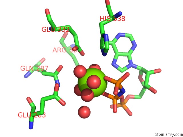
Mono view
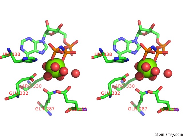
Stereo pair view
Mono view
Stereo pair view
A full contact list of Magnesium with other atoms in the Mg binding
site number 1 of Crystal Structure of the Catalytic Domain of Mmomers, An O-Methyl Tyrosyl-Trna Synthetase Evolved From Methanosarcina Mazei Pylrs, Complexed with O-Methyl Tyrosine and Amp-Pnp within 5.0Å range:
|
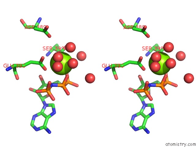
Magnesium binding site 2 out of 2 in 3qtc
Go back to
Magnesium binding site 2 out
of 2 in the Crystal Structure of the Catalytic Domain of Mmomers, An O-Methyl Tyrosyl-Trna Synthetase Evolved From Methanosarcina Mazei Pylrs, Complexed with O-Methyl Tyrosine and Amp-Pnp
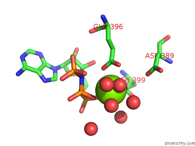
Mono view
Stereo pair view
Mono view
Stereo pair view
A full contact list of Magnesium with other atoms in the Mg binding
site number 2 of Crystal Structure of the Catalytic Domain of Mmomers, An O-Methyl Tyrosyl-Trna Synthetase Evolved From Methanosarcina Mazei Pylrs, Complexed with O-Methyl Tyrosine and Amp-Pnp within 5.0Å range:
|
Reference:
J.K.Takimoto,
N.Dellas,
J.P.Noel,
L.Wang.
Stereochemical Basis For Engineered Pyrrolysyl-Trna Synthetase and the Efficient in Vivo Incorporation of Structurally Divergent Non-Native Amino Acids. Acs Chem.Biol. V. 6 733 2011.
ISSN: ISSN 1554-8929
PubMed: 21545173
DOI: 10.1021/CB200057A
Page generated: Thu Aug 15 10:09:38 2024
ISSN: ISSN 1554-8929
PubMed: 21545173
DOI: 10.1021/CB200057A
Last articles
Zn in 9JYWZn in 9IR4
Zn in 9IR3
Zn in 9GMX
Zn in 9GMW
Zn in 9JEJ
Zn in 9ERF
Zn in 9ERE
Zn in 9EGV
Zn in 9EGW