Magnesium »
PDB 3qpp-3r10 »
3qtp »
Magnesium in PDB 3qtp: Crystal Structure Analysis of Entamoeba Histolytica Enolase
Enzymatic activity of Crystal Structure Analysis of Entamoeba Histolytica Enolase
All present enzymatic activity of Crystal Structure Analysis of Entamoeba Histolytica Enolase:
4.2.1.11;
4.2.1.11;
Protein crystallography data
The structure of Crystal Structure Analysis of Entamoeba Histolytica Enolase, PDB code: 3qtp
was solved by
E.C.Schulz,
R.Ficner,
with X-Ray Crystallography technique. A brief refinement statistics is given in the table below:
| Resolution Low / High (Å) | 46.31 / 1.90 |
| Space group | P 21 21 21 |
| Cell size a, b, c (Å), α, β, γ (°) | 64.420, 92.620, 160.580, 90.00, 90.00, 90.00 |
| R / Rfree (%) | 15.3 / 20.2 |
Magnesium Binding Sites:
The binding sites of Magnesium atom in the Crystal Structure Analysis of Entamoeba Histolytica Enolase
(pdb code 3qtp). This binding sites where shown within
5.0 Angstroms radius around Magnesium atom.
In total 2 binding sites of Magnesium where determined in the Crystal Structure Analysis of Entamoeba Histolytica Enolase, PDB code: 3qtp:
Jump to Magnesium binding site number: 1; 2;
In total 2 binding sites of Magnesium where determined in the Crystal Structure Analysis of Entamoeba Histolytica Enolase, PDB code: 3qtp:
Jump to Magnesium binding site number: 1; 2;
Magnesium binding site 1 out of 2 in 3qtp
Go back to
Magnesium binding site 1 out
of 2 in the Crystal Structure Analysis of Entamoeba Histolytica Enolase
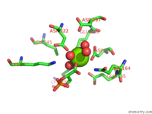
Mono view
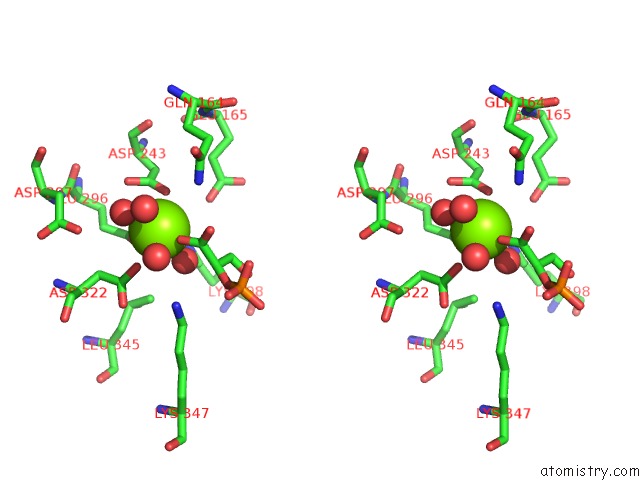
Stereo pair view
Mono view
Stereo pair view
A full contact list of Magnesium with other atoms in the Mg binding
site number 1 of Crystal Structure Analysis of Entamoeba Histolytica Enolase within 5.0Å range:
|
Magnesium binding site 2 out of 2 in 3qtp
Go back to
Magnesium binding site 2 out
of 2 in the Crystal Structure Analysis of Entamoeba Histolytica Enolase
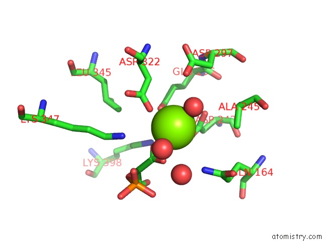
Mono view
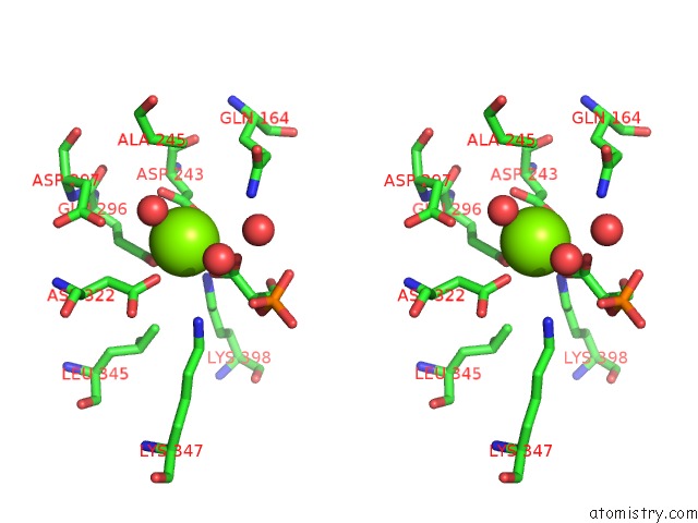
Stereo pair view
Mono view
Stereo pair view
A full contact list of Magnesium with other atoms in the Mg binding
site number 2 of Crystal Structure Analysis of Entamoeba Histolytica Enolase within 5.0Å range:
|
Reference:
E.C.Schulz,
M.Tietzel,
A.Tovy,
S.Ankri,
R.Ficner.
Structure Analysis of Entamoeba Histolytica Enolase. Acta Crystallogr.,Sect.D V. 67 619 2011.
ISSN: ISSN 0907-4449
PubMed: 21697600
DOI: 10.1107/S0907444911016544
Page generated: Thu Aug 15 10:10:17 2024
ISSN: ISSN 0907-4449
PubMed: 21697600
DOI: 10.1107/S0907444911016544
Last articles
Zn in 9JYWZn in 9IR4
Zn in 9IR3
Zn in 9GMX
Zn in 9GMW
Zn in 9JEJ
Zn in 9ERF
Zn in 9ERE
Zn in 9EGV
Zn in 9EGW