Magnesium »
PDB 3qpp-3r10 »
3qxh »
Magnesium in PDB 3qxh: Crystal Structure of Dethiobiotin Synthetase (Biod) From Helicobacter Pylori Complexed with Adp and 8-Aminocaprylic Acid
Enzymatic activity of Crystal Structure of Dethiobiotin Synthetase (Biod) From Helicobacter Pylori Complexed with Adp and 8-Aminocaprylic Acid
All present enzymatic activity of Crystal Structure of Dethiobiotin Synthetase (Biod) From Helicobacter Pylori Complexed with Adp and 8-Aminocaprylic Acid:
6.3.3.3;
6.3.3.3;
Protein crystallography data
The structure of Crystal Structure of Dethiobiotin Synthetase (Biod) From Helicobacter Pylori Complexed with Adp and 8-Aminocaprylic Acid, PDB code: 3qxh
was solved by
P.J.Porebski,
M.M.Klimecka,
M.Chruszcz,
K.Murzyn,
C.Minor,
A.Joachimiak,
W.Minor,
Midwest Center For Structural Genomics (Mcsg),
with X-Ray Crystallography technique. A brief refinement statistics is given in the table below:
| Resolution Low / High (Å) | 50.00 / 1.36 |
| Space group | C 1 2 1 |
| Cell size a, b, c (Å), α, β, γ (°) | 81.526, 37.967, 68.795, 90.00, 101.15, 90.00 |
| R / Rfree (%) | 11.7 / 14.2 |
Magnesium Binding Sites:
The binding sites of Magnesium atom in the Crystal Structure of Dethiobiotin Synthetase (Biod) From Helicobacter Pylori Complexed with Adp and 8-Aminocaprylic Acid
(pdb code 3qxh). This binding sites where shown within
5.0 Angstroms radius around Magnesium atom.
In total 2 binding sites of Magnesium where determined in the Crystal Structure of Dethiobiotin Synthetase (Biod) From Helicobacter Pylori Complexed with Adp and 8-Aminocaprylic Acid, PDB code: 3qxh:
Jump to Magnesium binding site number: 1; 2;
In total 2 binding sites of Magnesium where determined in the Crystal Structure of Dethiobiotin Synthetase (Biod) From Helicobacter Pylori Complexed with Adp and 8-Aminocaprylic Acid, PDB code: 3qxh:
Jump to Magnesium binding site number: 1; 2;
Magnesium binding site 1 out of 2 in 3qxh
Go back to
Magnesium binding site 1 out
of 2 in the Crystal Structure of Dethiobiotin Synthetase (Biod) From Helicobacter Pylori Complexed with Adp and 8-Aminocaprylic Acid
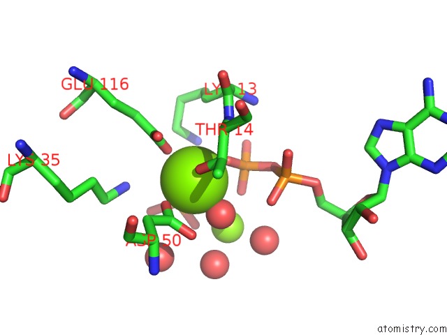
Mono view
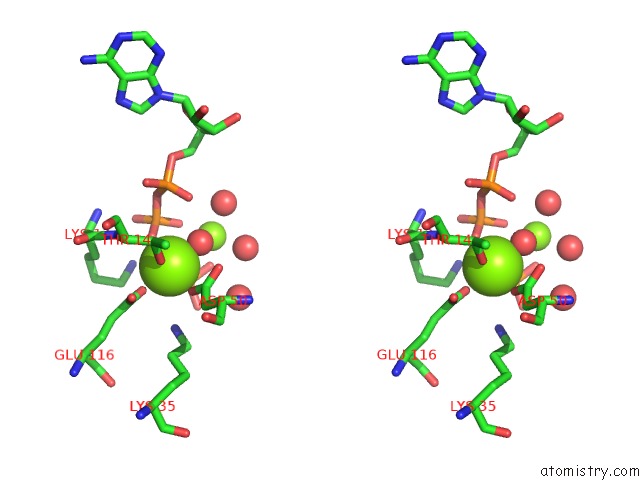
Stereo pair view
Mono view
Stereo pair view
A full contact list of Magnesium with other atoms in the Mg binding
site number 1 of Crystal Structure of Dethiobiotin Synthetase (Biod) From Helicobacter Pylori Complexed with Adp and 8-Aminocaprylic Acid within 5.0Å range:
|
Magnesium binding site 2 out of 2 in 3qxh
Go back to
Magnesium binding site 2 out
of 2 in the Crystal Structure of Dethiobiotin Synthetase (Biod) From Helicobacter Pylori Complexed with Adp and 8-Aminocaprylic Acid
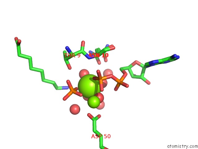
Mono view
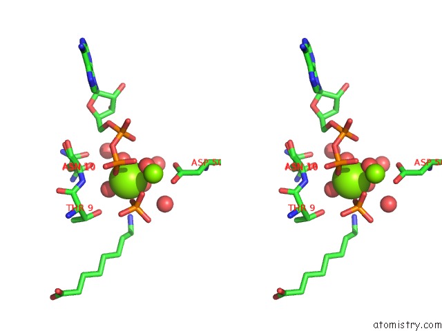
Stereo pair view
Mono view
Stereo pair view
A full contact list of Magnesium with other atoms in the Mg binding
site number 2 of Crystal Structure of Dethiobiotin Synthetase (Biod) From Helicobacter Pylori Complexed with Adp and 8-Aminocaprylic Acid within 5.0Å range:
|
Reference:
P.J.Porebski,
M.Klimecka,
M.Chruszcz,
R.A.Nicholls,
K.Murzyn,
M.E.Cuff,
X.Xu,
M.Cymborowski,
G.N.Murshudov,
A.Savchenko,
A.Edwards,
W.Minor.
Structural Characterization of Helicobacter Pylori Dethiobiotin Synthetase Reveals Differences Between Family Members. Febs J. V. 279 1093 2012.
ISSN: ISSN 1742-464X
PubMed: 22284390
DOI: 10.1111/J.1742-4658.2012.08506.X
Page generated: Thu Aug 15 10:12:47 2024
ISSN: ISSN 1742-464X
PubMed: 22284390
DOI: 10.1111/J.1742-4658.2012.08506.X
Last articles
Zn in 9JYWZn in 9IR4
Zn in 9IR3
Zn in 9GMX
Zn in 9GMW
Zn in 9JEJ
Zn in 9ERF
Zn in 9ERE
Zn in 9EGV
Zn in 9EGW