Magnesium »
PDB 3qpp-3r10 »
3qxr »
Magnesium in PDB 3qxr: Crystal Structure of the Brominated Ckit-1 Proto-Oncogene Promoter Quadruplex Dna
Protein crystallography data
The structure of Crystal Structure of the Brominated Ckit-1 Proto-Oncogene Promoter Quadruplex Dna, PDB code: 3qxr
was solved by
D.Wei,
G.N.Parkinson,
S.Neidle,
with X-Ray Crystallography technique. A brief refinement statistics is given in the table below:
| Resolution Low / High (Å) | 26.90 / 1.62 |
| Space group | P 21 21 2 |
| Cell size a, b, c (Å), α, β, γ (°) | 37.570, 51.350, 58.340, 90.00, 90.00, 90.00 |
| R / Rfree (%) | 16.6 / 21.3 |
Other elements in 3qxr:
The structure of Crystal Structure of the Brominated Ckit-1 Proto-Oncogene Promoter Quadruplex Dna also contains other interesting chemical elements:
| Bromine | (Br) | 2 atoms |
| Potassium | (K) | 9 atoms |
Magnesium Binding Sites:
The binding sites of Magnesium atom in the Crystal Structure of the Brominated Ckit-1 Proto-Oncogene Promoter Quadruplex Dna
(pdb code 3qxr). This binding sites where shown within
5.0 Angstroms radius around Magnesium atom.
In total 2 binding sites of Magnesium where determined in the Crystal Structure of the Brominated Ckit-1 Proto-Oncogene Promoter Quadruplex Dna, PDB code: 3qxr:
Jump to Magnesium binding site number: 1; 2;
In total 2 binding sites of Magnesium where determined in the Crystal Structure of the Brominated Ckit-1 Proto-Oncogene Promoter Quadruplex Dna, PDB code: 3qxr:
Jump to Magnesium binding site number: 1; 2;
Magnesium binding site 1 out of 2 in 3qxr
Go back to
Magnesium binding site 1 out
of 2 in the Crystal Structure of the Brominated Ckit-1 Proto-Oncogene Promoter Quadruplex Dna
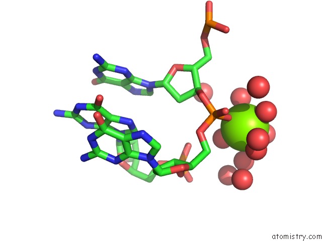
Mono view
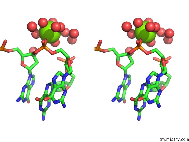
Stereo pair view
Mono view
Stereo pair view
A full contact list of Magnesium with other atoms in the Mg binding
site number 1 of Crystal Structure of the Brominated Ckit-1 Proto-Oncogene Promoter Quadruplex Dna within 5.0Å range:
|
Magnesium binding site 2 out of 2 in 3qxr
Go back to
Magnesium binding site 2 out
of 2 in the Crystal Structure of the Brominated Ckit-1 Proto-Oncogene Promoter Quadruplex Dna
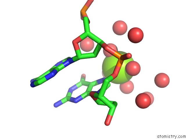
Mono view
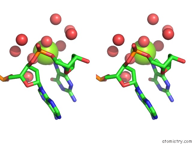
Stereo pair view
Mono view
Stereo pair view
A full contact list of Magnesium with other atoms in the Mg binding
site number 2 of Crystal Structure of the Brominated Ckit-1 Proto-Oncogene Promoter Quadruplex Dna within 5.0Å range:
|
Reference:
D.Wei,
G.N.Parkinson,
A.P.Reszka,
S.Neidle.
Crystal Structure of A C-Kit Promoter Quadruplex Reveals the Structural Role of Metal Ions and Water Molecules in Maintaining Loop Conformation. Nucleic Acids Res. V. 40 4691 2012.
ISSN: ISSN 0305-1048
PubMed: 22287624
DOI: 10.1093/NAR/GKS023
Page generated: Thu Aug 15 10:12:52 2024
ISSN: ISSN 0305-1048
PubMed: 22287624
DOI: 10.1093/NAR/GKS023
Last articles
Zn in 9J0NZn in 9J0O
Zn in 9J0P
Zn in 9FJX
Zn in 9EKB
Zn in 9C0F
Zn in 9CAH
Zn in 9CH0
Zn in 9CH3
Zn in 9CH1