Magnesium »
PDB 3r11-3ren »
3rab »
Magnesium in PDB 3rab: Gppnhp-Bound RAB3A at 2.0 A Resolution
Protein crystallography data
The structure of Gppnhp-Bound RAB3A at 2.0 A Resolution, PDB code: 3rab
was solved by
J.J.Dumas,
Z.Zhu,
J.L.Connolly,
D.G.Lambright,
with X-Ray Crystallography technique. A brief refinement statistics is given in the table below:
| Resolution Low / High (Å) | 8.00 / 2.00 |
| Space group | C 1 2 1 |
| Cell size a, b, c (Å), α, β, γ (°) | 88.900, 35.000, 58.600, 90.00, 107.20, 90.00 |
| R / Rfree (%) | 19.1 / 23.7 |
Magnesium Binding Sites:
The binding sites of Magnesium atom in the Gppnhp-Bound RAB3A at 2.0 A Resolution
(pdb code 3rab). This binding sites where shown within
5.0 Angstroms radius around Magnesium atom.
In total only one binding site of Magnesium was determined in the Gppnhp-Bound RAB3A at 2.0 A Resolution, PDB code: 3rab:
In total only one binding site of Magnesium was determined in the Gppnhp-Bound RAB3A at 2.0 A Resolution, PDB code: 3rab:
Magnesium binding site 1 out of 1 in 3rab
Go back to
Magnesium binding site 1 out
of 1 in the Gppnhp-Bound RAB3A at 2.0 A Resolution
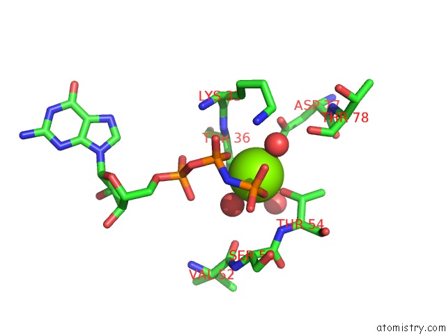
Mono view
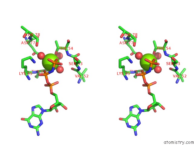
Stereo pair view
Mono view
Stereo pair view
A full contact list of Magnesium with other atoms in the Mg binding
site number 1 of Gppnhp-Bound RAB3A at 2.0 A Resolution within 5.0Å range:
|
Reference:
J.J.Dumas,
Z.Zhu,
J.L.Connolly,
D.G.Lambright.
Structural Basis of Activation and Gtp Hydrolysis in Rab Proteins. Structure Fold.Des. V. 7 413 1999.
ISSN: ISSN 0969-2126
PubMed: 10196122
DOI: 10.1016/S0969-2126(99)80054-9
Page generated: Thu Aug 15 10:21:38 2024
ISSN: ISSN 0969-2126
PubMed: 10196122
DOI: 10.1016/S0969-2126(99)80054-9
Last articles
Ca in 5SB8Ca in 5SB6
Ca in 5SB5
Ca in 5SB3
Ca in 5SB4
Ca in 5S9M
Ca in 5S9N
Ca in 5S9L
Ca in 5S8Q
Ca in 5S8P