Magnesium »
PDB 3rer-3rqx »
3rq6 »
Magnesium in PDB 3rq6: Crystal Structure of Adp/Atp-Dependent Nad(P)H-Hydrate Dehydratase From Bacillus Subtilis Soaked with Adp-Ribose
Enzymatic activity of Crystal Structure of Adp/Atp-Dependent Nad(P)H-Hydrate Dehydratase From Bacillus Subtilis Soaked with Adp-Ribose
All present enzymatic activity of Crystal Structure of Adp/Atp-Dependent Nad(P)H-Hydrate Dehydratase From Bacillus Subtilis Soaked with Adp-Ribose:
4.2.1.93;
4.2.1.93;
Protein crystallography data
The structure of Crystal Structure of Adp/Atp-Dependent Nad(P)H-Hydrate Dehydratase From Bacillus Subtilis Soaked with Adp-Ribose, PDB code: 3rq6
was solved by
I.A.Shumilin,
M.Cymborowski,
A.Joachimiak,
W.Minor,
Midwest Center Forstructural Genomics (Mcsg),
with X-Ray Crystallography technique. A brief refinement statistics is given in the table below:
| Resolution Low / High (Å) | 50.00 / 1.65 |
| Space group | I 4 2 2 |
| Cell size a, b, c (Å), α, β, γ (°) | 91.542, 91.542, 169.131, 90.00, 90.00, 90.00 |
| R / Rfree (%) | 15 / 17.6 |
Magnesium Binding Sites:
The binding sites of Magnesium atom in the Crystal Structure of Adp/Atp-Dependent Nad(P)H-Hydrate Dehydratase From Bacillus Subtilis Soaked with Adp-Ribose
(pdb code 3rq6). This binding sites where shown within
5.0 Angstroms radius around Magnesium atom.
In total only one binding site of Magnesium was determined in the Crystal Structure of Adp/Atp-Dependent Nad(P)H-Hydrate Dehydratase From Bacillus Subtilis Soaked with Adp-Ribose, PDB code: 3rq6:
In total only one binding site of Magnesium was determined in the Crystal Structure of Adp/Atp-Dependent Nad(P)H-Hydrate Dehydratase From Bacillus Subtilis Soaked with Adp-Ribose, PDB code: 3rq6:
Magnesium binding site 1 out of 1 in 3rq6
Go back to
Magnesium binding site 1 out
of 1 in the Crystal Structure of Adp/Atp-Dependent Nad(P)H-Hydrate Dehydratase From Bacillus Subtilis Soaked with Adp-Ribose
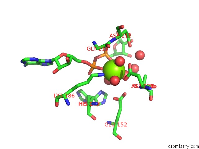
Mono view
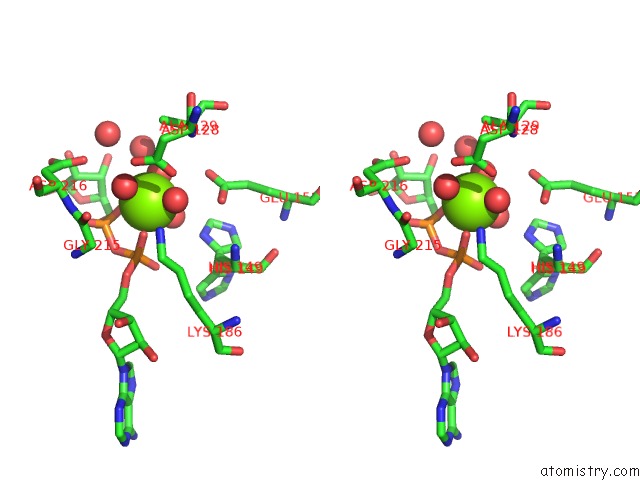
Stereo pair view
Mono view
Stereo pair view
A full contact list of Magnesium with other atoms in the Mg binding
site number 1 of Crystal Structure of Adp/Atp-Dependent Nad(P)H-Hydrate Dehydratase From Bacillus Subtilis Soaked with Adp-Ribose within 5.0Å range:
|
Reference:
I.A.Shumilin,
M.Cymborowski,
O.Chertihin,
K.N.Jha,
J.C.Herr,
S.A.Lesley,
A.Joachimiak,
W.Minor.
Identification of Unknown Protein Function Using Metabolite Cocktail Screening. Structure V. 20 1715 2012.
ISSN: ISSN 0969-2126
PubMed: 22940582
DOI: 10.1016/J.STR.2012.07.016
Page generated: Thu Aug 15 10:33:48 2024
ISSN: ISSN 0969-2126
PubMed: 22940582
DOI: 10.1016/J.STR.2012.07.016
Last articles
Zn in 9J0NZn in 9J0O
Zn in 9J0P
Zn in 9FJX
Zn in 9EKB
Zn in 9C0F
Zn in 9CAH
Zn in 9CH0
Zn in 9CH3
Zn in 9CH1