Magnesium »
PDB 3rr7-3rv6 »
3rtv »
Magnesium in PDB 3rtv: Crystal Structure of the Large Fragment of Dna Polymerase I From Thermus Aquaticus in A Closed Ternary Complex with Natural Primer/Template Dna
Enzymatic activity of Crystal Structure of the Large Fragment of Dna Polymerase I From Thermus Aquaticus in A Closed Ternary Complex with Natural Primer/Template Dna
All present enzymatic activity of Crystal Structure of the Large Fragment of Dna Polymerase I From Thermus Aquaticus in A Closed Ternary Complex with Natural Primer/Template Dna:
2.7.7.7;
2.7.7.7;
Protein crystallography data
The structure of Crystal Structure of the Large Fragment of Dna Polymerase I From Thermus Aquaticus in A Closed Ternary Complex with Natural Primer/Template Dna, PDB code: 3rtv
was solved by
A.Marx,
K.Diederichs,
K.Betz,
with X-Ray Crystallography technique. A brief refinement statistics is given in the table below:
| Resolution Low / High (Å) | 46.89 / 1.90 |
| Space group | P 31 2 1 |
| Cell size a, b, c (Å), α, β, γ (°) | 108.296, 108.296, 90.381, 90.00, 90.00, 120.00 |
| R / Rfree (%) | 16.2 / 20.2 |
Magnesium Binding Sites:
The binding sites of Magnesium atom in the Crystal Structure of the Large Fragment of Dna Polymerase I From Thermus Aquaticus in A Closed Ternary Complex with Natural Primer/Template Dna
(pdb code 3rtv). This binding sites where shown within
5.0 Angstroms radius around Magnesium atom.
In total 3 binding sites of Magnesium where determined in the Crystal Structure of the Large Fragment of Dna Polymerase I From Thermus Aquaticus in A Closed Ternary Complex with Natural Primer/Template Dna, PDB code: 3rtv:
Jump to Magnesium binding site number: 1; 2; 3;
In total 3 binding sites of Magnesium where determined in the Crystal Structure of the Large Fragment of Dna Polymerase I From Thermus Aquaticus in A Closed Ternary Complex with Natural Primer/Template Dna, PDB code: 3rtv:
Jump to Magnesium binding site number: 1; 2; 3;
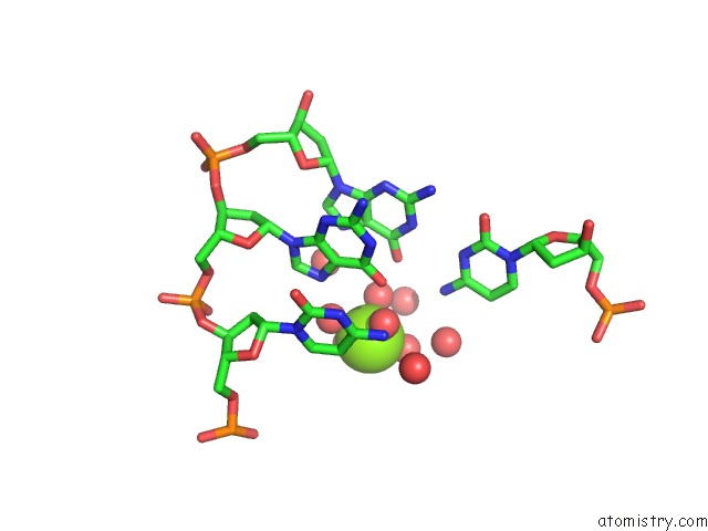
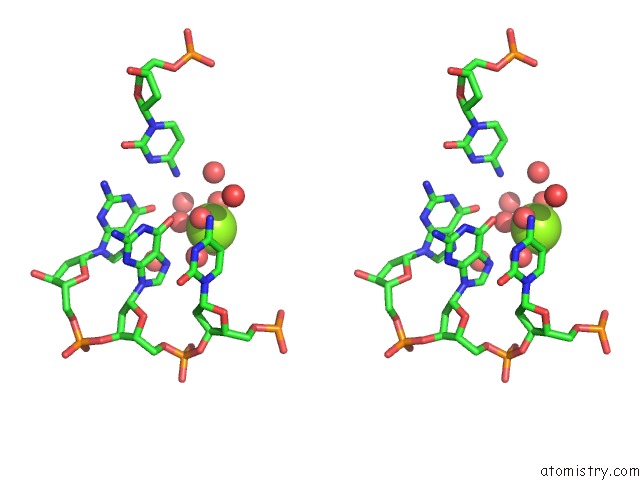
Magnesium binding site 1 out of 3 in 3rtv
Go back to
Magnesium binding site 1 out
of 3 in the Crystal Structure of the Large Fragment of Dna Polymerase I From Thermus Aquaticus in A Closed Ternary Complex with Natural Primer/Template Dna
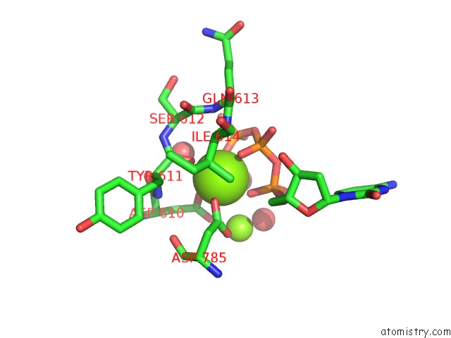
Mono view
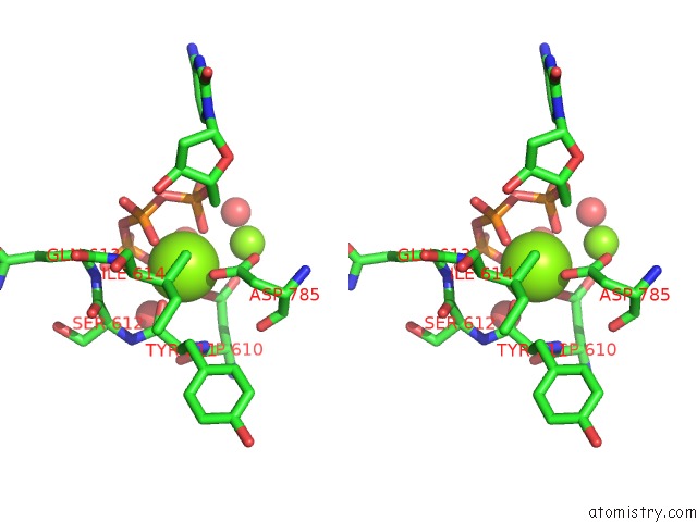
Stereo pair view
Mono view
Stereo pair view
A full contact list of Magnesium with other atoms in the Mg binding
site number 1 of Crystal Structure of the Large Fragment of Dna Polymerase I From Thermus Aquaticus in A Closed Ternary Complex with Natural Primer/Template Dna within 5.0Å range:
|
Magnesium binding site 2 out of 3 in 3rtv
Go back to
Magnesium binding site 2 out
of 3 in the Crystal Structure of the Large Fragment of Dna Polymerase I From Thermus Aquaticus in A Closed Ternary Complex with Natural Primer/Template Dna
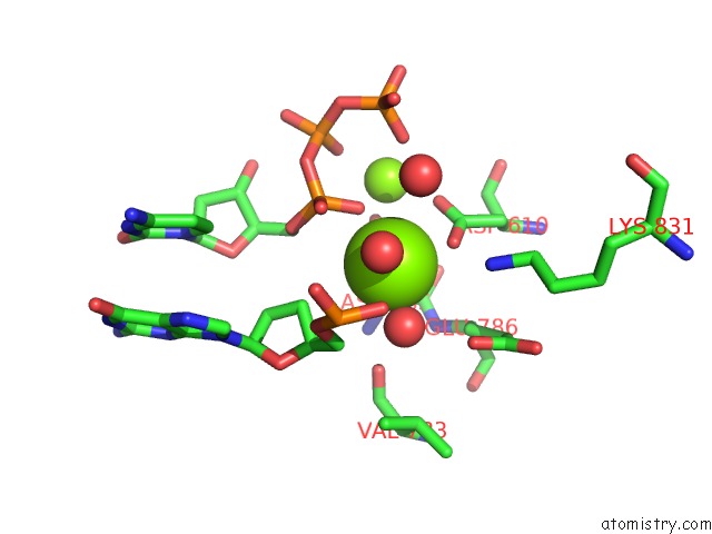
Mono view
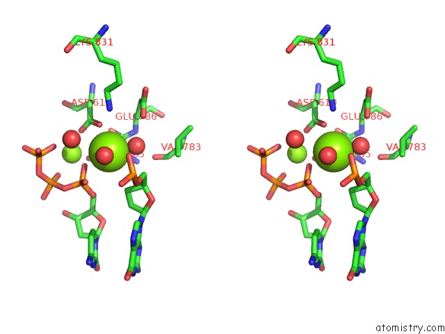
Stereo pair view
Mono view
Stereo pair view
A full contact list of Magnesium with other atoms in the Mg binding
site number 2 of Crystal Structure of the Large Fragment of Dna Polymerase I From Thermus Aquaticus in A Closed Ternary Complex with Natural Primer/Template Dna within 5.0Å range:
|
Magnesium binding site 3 out of 3 in 3rtv
Go back to
Magnesium binding site 3 out
of 3 in the Crystal Structure of the Large Fragment of Dna Polymerase I From Thermus Aquaticus in A Closed Ternary Complex with Natural Primer/Template Dna
Mono view
Stereo pair view
Mono view
Stereo pair view
A full contact list of Magnesium with other atoms in the Mg binding
site number 3 of Crystal Structure of the Large Fragment of Dna Polymerase I From Thermus Aquaticus in A Closed Ternary Complex with Natural Primer/Template Dna within 5.0Å range:
|
Reference:
K.Betz,
D.A.Malyshev,
T.Lavergne,
W.Welte,
K.Diederichs,
T.J.Dwyer,
P.Ordoukhanian,
F.E.Romesberg,
A.Marx.
Klentaq Polymerase Replicates Unnatural Base Pairs By Inducing A Watson-Crick Geometry. Nat.Chem.Biol. V. 8 612 2012.
ISSN: ISSN 1552-4450
PubMed: 22660438
DOI: 10.1038/NCHEMBIO.966
Page generated: Thu Aug 15 10:38:47 2024
ISSN: ISSN 1552-4450
PubMed: 22660438
DOI: 10.1038/NCHEMBIO.966
Last articles
Zn in 9MJ5Zn in 9HNW
Zn in 9G0L
Zn in 9FNE
Zn in 9DZN
Zn in 9E0I
Zn in 9D32
Zn in 9DAK
Zn in 8ZXC
Zn in 8ZUF