Magnesium »
PDB 3rr7-3rv6 »
3rv3 »
Magnesium in PDB 3rv3: Crystal Structure of E.Coli Biotin Carboxylase in Complex with Two Adp and One Mg Ion
Enzymatic activity of Crystal Structure of E.Coli Biotin Carboxylase in Complex with Two Adp and One Mg Ion
All present enzymatic activity of Crystal Structure of E.Coli Biotin Carboxylase in Complex with Two Adp and One Mg Ion:
6.3.4.14; 6.4.1.2;
6.3.4.14; 6.4.1.2;
Protein crystallography data
The structure of Crystal Structure of E.Coli Biotin Carboxylase in Complex with Two Adp and One Mg Ion, PDB code: 3rv3
was solved by
C.Y.Chou,
L.Tong,
with X-Ray Crystallography technique. A brief refinement statistics is given in the table below:
| Resolution Low / High (Å) | 30.00 / 1.91 |
| Space group | C 1 2 1 |
| Cell size a, b, c (Å), α, β, γ (°) | 171.357, 58.009, 85.236, 90.00, 94.64, 90.00 |
| R / Rfree (%) | 20.8 / 26.3 |
Magnesium Binding Sites:
The binding sites of Magnesium atom in the Crystal Structure of E.Coli Biotin Carboxylase in Complex with Two Adp and One Mg Ion
(pdb code 3rv3). This binding sites where shown within
5.0 Angstroms radius around Magnesium atom.
In total 2 binding sites of Magnesium where determined in the Crystal Structure of E.Coli Biotin Carboxylase in Complex with Two Adp and One Mg Ion, PDB code: 3rv3:
Jump to Magnesium binding site number: 1; 2;
In total 2 binding sites of Magnesium where determined in the Crystal Structure of E.Coli Biotin Carboxylase in Complex with Two Adp and One Mg Ion, PDB code: 3rv3:
Jump to Magnesium binding site number: 1; 2;
Magnesium binding site 1 out of 2 in 3rv3
Go back to
Magnesium binding site 1 out
of 2 in the Crystal Structure of E.Coli Biotin Carboxylase in Complex with Two Adp and One Mg Ion
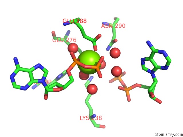
Mono view
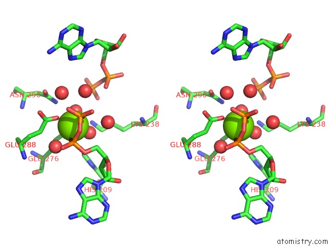
Stereo pair view
Mono view
Stereo pair view
A full contact list of Magnesium with other atoms in the Mg binding
site number 1 of Crystal Structure of E.Coli Biotin Carboxylase in Complex with Two Adp and One Mg Ion within 5.0Å range:
|
Magnesium binding site 2 out of 2 in 3rv3
Go back to
Magnesium binding site 2 out
of 2 in the Crystal Structure of E.Coli Biotin Carboxylase in Complex with Two Adp and One Mg Ion
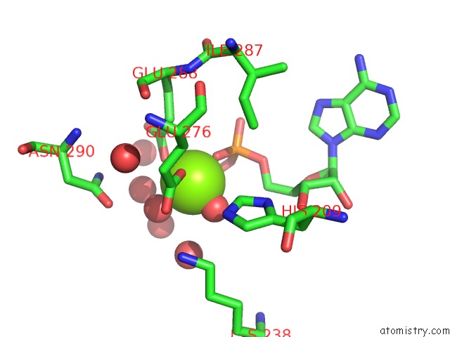
Mono view
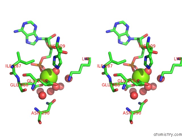
Stereo pair view
Mono view
Stereo pair view
A full contact list of Magnesium with other atoms in the Mg binding
site number 2 of Crystal Structure of E.Coli Biotin Carboxylase in Complex with Two Adp and One Mg Ion within 5.0Å range:
|
Reference:
C.Y.Chou,
L.Tong.
Structural and Biochemical Studies on the Regulation of Biotin Carboxylase By Substrate Inhibition and Dimerization. J.Biol.Chem. V. 286 24417 2011.
ISSN: ISSN 0021-9258
PubMed: 21592965
DOI: 10.1074/JBC.M111.220517
Page generated: Thu Aug 15 10:40:32 2024
ISSN: ISSN 0021-9258
PubMed: 21592965
DOI: 10.1074/JBC.M111.220517
Last articles
Zn in 9MJ5Zn in 9HNW
Zn in 9G0L
Zn in 9FNE
Zn in 9DZN
Zn in 9E0I
Zn in 9D32
Zn in 9DAK
Zn in 8ZXC
Zn in 8ZUF