Magnesium »
PDB 3rv6-3s8b »
3rv6 »
Magnesium in PDB 3rv6: Structure of A M. Tuberculosis Salicylate Synthase, Mbti, in Complex with An Inhibitor with Phenyl R-Group
Enzymatic activity of Structure of A M. Tuberculosis Salicylate Synthase, Mbti, in Complex with An Inhibitor with Phenyl R-Group
All present enzymatic activity of Structure of A M. Tuberculosis Salicylate Synthase, Mbti, in Complex with An Inhibitor with Phenyl R-Group:
5.4.4.2;
5.4.4.2;
Protein crystallography data
The structure of Structure of A M. Tuberculosis Salicylate Synthase, Mbti, in Complex with An Inhibitor with Phenyl R-Group, PDB code: 3rv6
was solved by
G.Chi,
E.M.M.Bulloch,
A.Manos-Turvey,
R.J.Payne,
J.S.Lott,
Tb Structuralgenomics Consortium (Tbsgc),
with X-Ray Crystallography technique. A brief refinement statistics is given in the table below:
| Resolution Low / High (Å) | 16.72 / 2.04 |
| Space group | P 1 21 1 |
| Cell size a, b, c (Å), α, β, γ (°) | 52.748, 91.530, 96.466, 90.00, 104.83, 90.00 |
| R / Rfree (%) | 17.1 / 20.5 |
Magnesium Binding Sites:
The binding sites of Magnesium atom in the Structure of A M. Tuberculosis Salicylate Synthase, Mbti, in Complex with An Inhibitor with Phenyl R-Group
(pdb code 3rv6). This binding sites where shown within
5.0 Angstroms radius around Magnesium atom.
In total 2 binding sites of Magnesium where determined in the Structure of A M. Tuberculosis Salicylate Synthase, Mbti, in Complex with An Inhibitor with Phenyl R-Group, PDB code: 3rv6:
Jump to Magnesium binding site number: 1; 2;
In total 2 binding sites of Magnesium where determined in the Structure of A M. Tuberculosis Salicylate Synthase, Mbti, in Complex with An Inhibitor with Phenyl R-Group, PDB code: 3rv6:
Jump to Magnesium binding site number: 1; 2;
Magnesium binding site 1 out of 2 in 3rv6
Go back to
Magnesium binding site 1 out
of 2 in the Structure of A M. Tuberculosis Salicylate Synthase, Mbti, in Complex with An Inhibitor with Phenyl R-Group
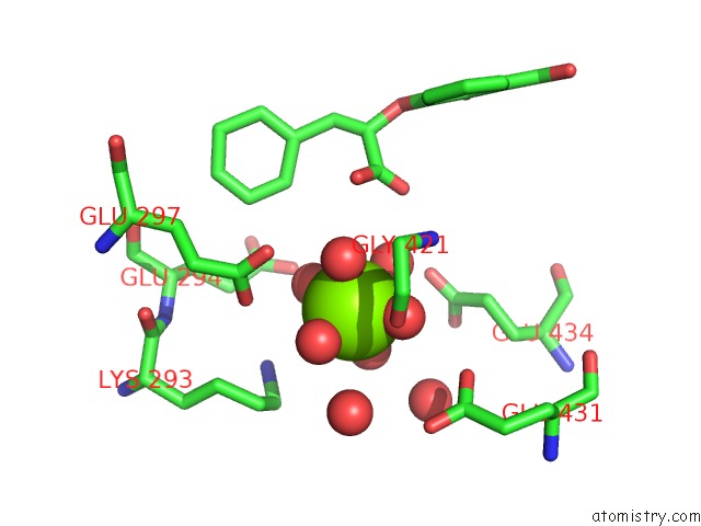
Mono view
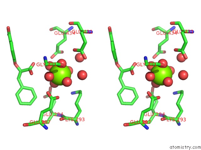
Stereo pair view
Mono view
Stereo pair view
A full contact list of Magnesium with other atoms in the Mg binding
site number 1 of Structure of A M. Tuberculosis Salicylate Synthase, Mbti, in Complex with An Inhibitor with Phenyl R-Group within 5.0Å range:
|
Magnesium binding site 2 out of 2 in 3rv6
Go back to
Magnesium binding site 2 out
of 2 in the Structure of A M. Tuberculosis Salicylate Synthase, Mbti, in Complex with An Inhibitor with Phenyl R-Group
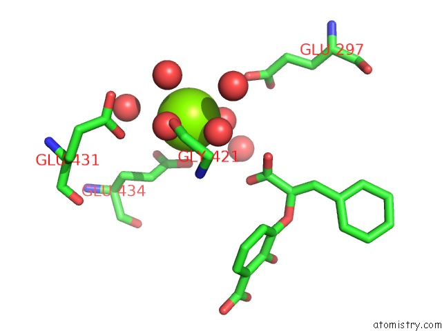
Mono view
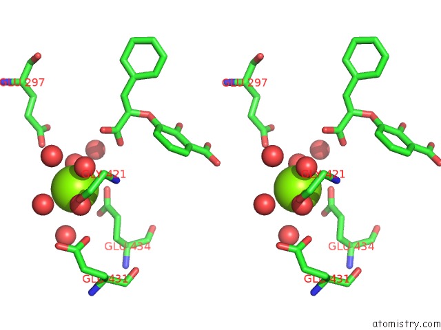
Stereo pair view
Mono view
Stereo pair view
A full contact list of Magnesium with other atoms in the Mg binding
site number 2 of Structure of A M. Tuberculosis Salicylate Synthase, Mbti, in Complex with An Inhibitor with Phenyl R-Group within 5.0Å range:
|
Reference:
G.Chi,
A.Manos-Turvey,
P.D.O'connor,
J.M.Johnston,
G.L.Evans,
E.N.Baker,
R.J.Payne,
J.S.Lott,
E.M.Bulloch.
Implications of Binding Mode and Active Site Flexibility For Inhibitor Potency Against the Salicylate Synthase From Mycobacterium Tuberculosis Biochemistry V. 51 4868 2012.
ISSN: ISSN 0006-2960
PubMed: 22607697
DOI: 10.1021/BI3002067
Page generated: Thu Aug 15 10:41:49 2024
ISSN: ISSN 0006-2960
PubMed: 22607697
DOI: 10.1021/BI3002067
Last articles
Zn in 9J0NZn in 9J0O
Zn in 9J0P
Zn in 9FJX
Zn in 9EKB
Zn in 9C0F
Zn in 9CAH
Zn in 9CH0
Zn in 9CH3
Zn in 9CH1