Atomistry » Magnesium » PDB 3rv6-3s8b » 3s1a
Atomistry »
  Magnesium »
    PDB 3rv6-3s8b »
      3s1a »

Magnesium in PDB 3s1a: Crystal Structure of the Phosphorylation-Site Double Mutant S431E/T432E of the Kaic Circadian Clock Protein

Enzymatic activity of Crystal Structure of the Phosphorylation-Site Double Mutant S431E/T432E of the Kaic Circadian Clock Protein

All present enzymatic activity of Crystal Structure of the Phosphorylation-Site Double Mutant S431E/T432E of the Kaic Circadian Clock Protein:
2.7.11.1;

Protein crystallography data

The structure of Crystal Structure of the Phosphorylation-Site Double Mutant S431E/T432E of the Kaic Circadian Clock Protein, PDB code: 3s1a was solved by R.Pattanayek, D.W.Williams, G.Rossi, S.Weigand, T.Mori, C.H.Johnson, P.L.Stewart, M.Egli, with X-Ray Crystallography technique. A brief refinement statistics is given in the table below:

Resolution Low / High (Å) 17.00 / 3.00
Space group P 21 21 21
Cell size a, b, c (Å), α, β, γ (°) 132.666, 135.494, 204.603, 90.00, 90.00, 90.00
R / Rfree (%) 24.2 / 28.8

Magnesium Binding Sites:

Pages:

>>> Page 1 <<< Page 2, Binding sites: 11 - 20; Page 3, Binding sites: 21 - 21;

Binding sites:

The binding sites of Magnesium atom in the Crystal Structure of the Phosphorylation-Site Double Mutant S431E/T432E of the Kaic Circadian Clock Protein (pdb code 3s1a). This binding sites where shown within 5.0 Angstroms radius around Magnesium atom.
In total 21 binding sites of Magnesium where determined in the Crystal Structure of the Phosphorylation-Site Double Mutant S431E/T432E of the Kaic Circadian Clock Protein, PDB code: 3s1a:
Jump to Magnesium binding site number: 1; 2; 3; 4; 5; 6; 7; 8; 9; 10;

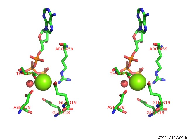
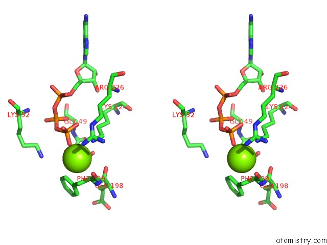
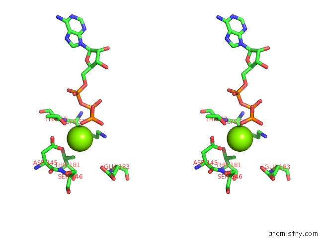
Magnesium binding site 1 out of 21 in 3s1a

Go back to Magnesium Binding Sites List in 3s1a
Magnesium binding site 1 out of 21 in the Crystal Structure of the Phosphorylation-Site Double Mutant S431E/T432E of the Kaic Circadian Clock Protein


Mono view


Stereo pair view

A full contact list of Magnesium with other atoms in the Mg binding site number 1 of Crystal Structure of the Phosphorylation-Site Double Mutant S431E/T432E of the Kaic Circadian Clock Protein within 5.0Å range:
probe atom residue distance (Å) B Occ
A:Mg801

b:46.1
occ:1.00
OG1 A:THR295 2.4 67.8 1.0
O A:HOH527 2.7 40.3 1.0
O2G A:ATP901 2.8 84.7 1.0
OE2 A:GLU319 2.9 95.8 1.0
O3B A:ATP901 3.0 84.7 1.0
PG A:ATP901 3.1 84.7 1.0
O3G A:ATP901 3.2 84.8 1.0
NH2 B:ARG459 3.5 81.4 1.0
O3A A:ATP901 3.7 85.7 1.0
OD2 A:ASP378 3.7 84.1 1.0
CB A:THR295 3.7 69.3 1.0
PB A:ATP901 4.0 84.7 1.0
CD A:GLU319 4.0 94.4 1.0
OE2 A:GLU318 4.0 97.4 1.0
NE B:ARG459 4.2 84.0 1.0
CZ B:ARG459 4.3 82.6 1.0
CG2 A:THR295 4.3 67.2 1.0
OD1 A:ASP378 4.4 81.5 1.0
CG A:ASP378 4.4 82.5 1.0
O1B A:ATP901 4.4 84.9 1.0
O2A A:ATP901 4.5 85.9 1.0
O1G A:ATP901 4.6 86.0 1.0
MG A:MG802 4.7 82.9 1.0
CD A:GLU318 4.7 96.6 1.0
OE1 A:GLU319 4.8 96.2 1.0
CA A:THR295 4.8 71.0 1.0
PA A:ATP901 4.8 86.1 1.0
CG A:GLU319 4.8 91.2 1.0
N A:THR295 4.9 71.9 1.0
CG A:GLU318 5.0 94.5 1.0

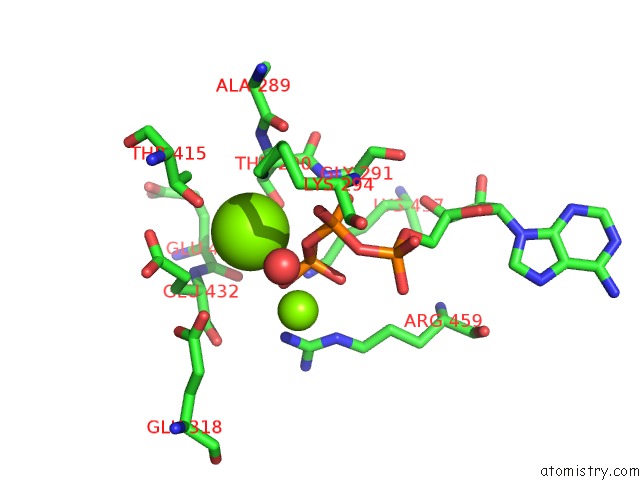
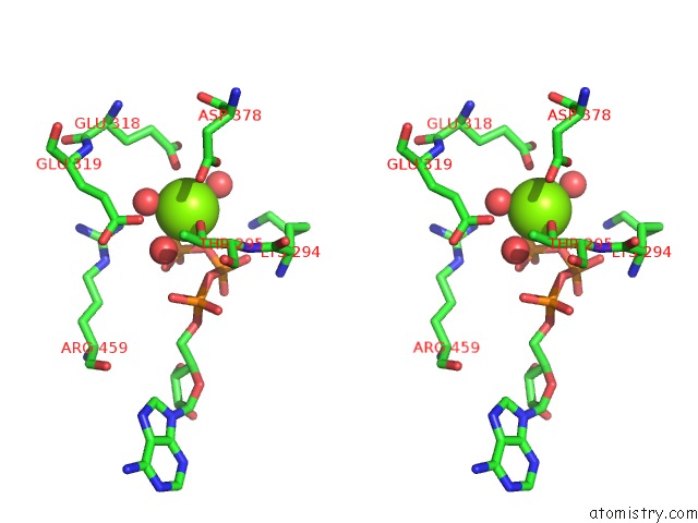
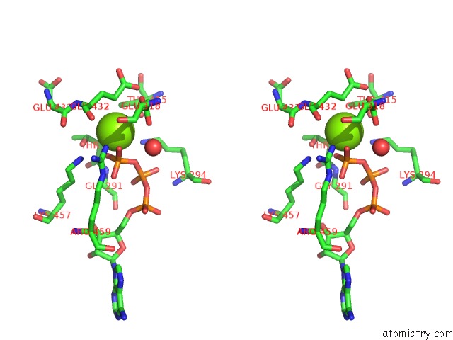
Magnesium binding site 2 out of 21 in 3s1a

Go back to Magnesium Binding Sites List in 3s1a
Magnesium binding site 2 out of 21 in the Crystal Structure of the Phosphorylation-Site Double Mutant S431E/T432E of the Kaic Circadian Clock Protein


Mono view


Stereo pair view

A full contact list of Magnesium with other atoms in the Mg binding site number 2 of Crystal Structure of the Phosphorylation-Site Double Mutant S431E/T432E of the Kaic Circadian Clock Protein within 5.0Å range:
probe atom residue distance (Å) B Occ
A:Mg802

b:82.9
occ:1.00
O2G A:ATP901 2.7 84.7 1.0
O1G A:ATP901 2.9 86.0 1.0
PG A:ATP901 3.0 84.7 1.0
O3B A:ATP901 3.0 84.7 1.0
OG1 A:THR415 3.3 60.3 1.0
NZ A:LYS294 3.4 82.7 1.0
CG B:GLU432 3.5 74.2 1.0
CA A:THR290 3.6 58.4 1.0
OG1 A:THR290 3.7 57.9 1.0
CG2 A:THR415 3.7 62.2 1.0
CB A:THR290 3.8 58.7 1.0
CB A:THR415 4.1 62.4 1.0
CD B:GLU432 4.2 76.1 1.0
CA B:GLU432 4.2 74.9 1.0
PB A:ATP901 4.3 84.7 1.0
N A:GLY291 4.3 60.9 1.0
CB B:GLU432 4.4 73.0 1.0
OE1 B:GLU432 4.4 77.1 1.0
NZ B:LYS457 4.4 71.0 1.0
O3G A:ATP901 4.5 84.8 1.0
C A:THR290 4.5 59.8 1.0
CE A:LYS294 4.5 81.5 1.0
OE2 A:GLU318 4.6 97.4 1.0
N A:THR290 4.6 57.1 1.0
MG A:MG801 4.7 46.1 1.0
O1B A:ATP901 4.7 84.9 1.0
O2B A:ATP901 4.7 85.0 1.0
N B:GLU432 4.8 72.4 1.0
O A:HOH527 4.8 40.3 1.0
O A:ALA289 4.9 57.9 1.0
O B:GLU431 5.0 72.7 1.0
NH2 B:ARG459 5.0 81.4 1.0

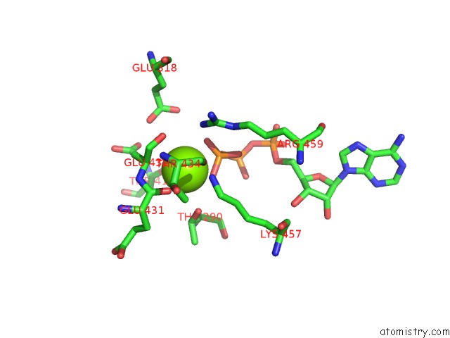
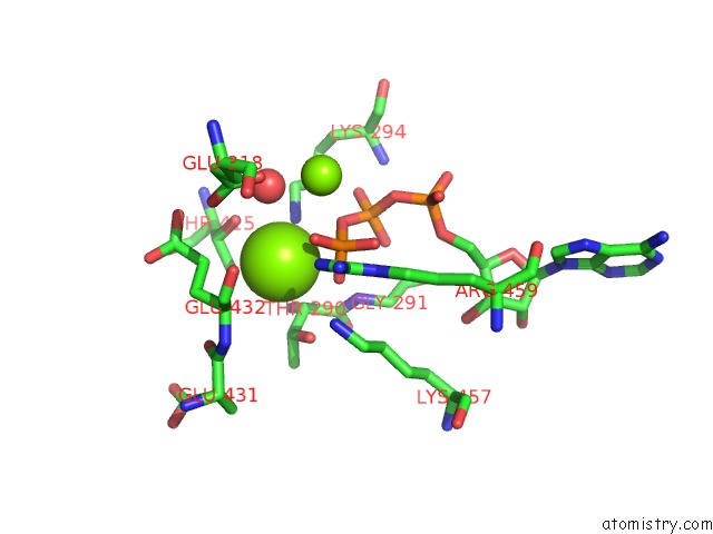
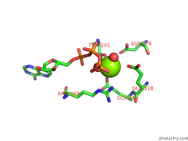
Magnesium binding site 3 out of 21 in 3s1a

Go back to Magnesium Binding Sites List in 3s1a
Magnesium binding site 3 out of 21 in the Crystal Structure of the Phosphorylation-Site Double Mutant S431E/T432E of the Kaic Circadian Clock Protein


Mono view


Stereo pair view

A full contact list of Magnesium with other atoms in the Mg binding site number 3 of Crystal Structure of the Phosphorylation-Site Double Mutant S431E/T432E of the Kaic Circadian Clock Protein within 5.0Å range:
probe atom residue distance (Å) B Occ
A:Mg701

b:63.9
occ:1.00
OG1 A:THR53 2.4 51.4 1.0
O2G A:ATP903 2.6 76.7 1.0
O3B A:ATP903 2.9 72.1 1.0
PG A:ATP903 3.1 78.0 1.0
OD2 A:ASP145 3.1 68.1 1.0
O1G A:ATP903 3.5 77.0 1.0
OD1 A:ASP145 3.7 65.9 1.0
N A:THR53 3.7 54.8 1.0
CB A:THR53 3.7 53.0 1.0
CG A:ASP145 3.8 65.7 1.0
CG2 A:THR181 3.8 50.9 1.0
CB A:LYS52 4.1 53.9 1.0
CA A:THR53 4.1 53.6 1.0
PB A:ATP903 4.2 66.2 1.0
CE A:LYS52 4.3 54.7 1.0
O3A A:ATP903 4.4 65.8 1.0
O1B A:ATP903 4.5 67.4 1.0
O3G A:ATP903 4.6 76.8 1.0
NZ A:LYS52 4.6 56.1 1.0
CG2 A:THR53 4.6 53.1 1.0
CD A:LYS52 4.7 55.1 1.0
C A:LYS52 4.7 54.8 1.0
OG1 A:THR181 4.9 51.8 1.0
CB A:THR181 4.9 51.5 1.0
CA A:LYS52 5.0 54.0 1.0
NH2 B:ARG226 5.0 74.8 1.0

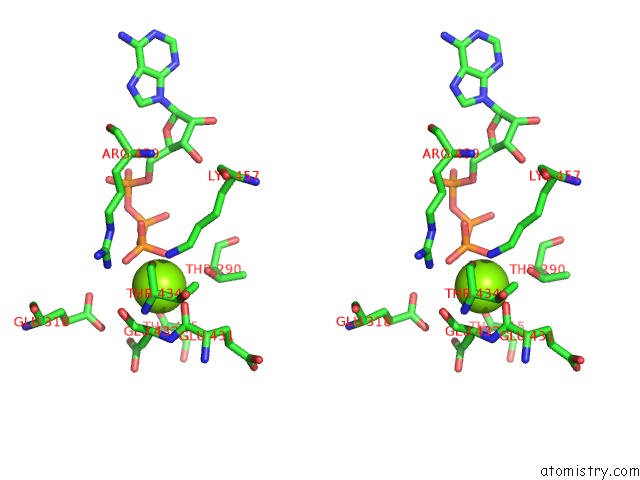
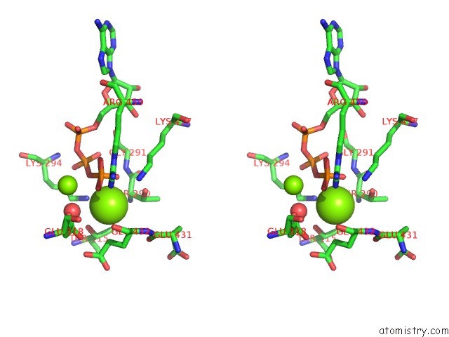
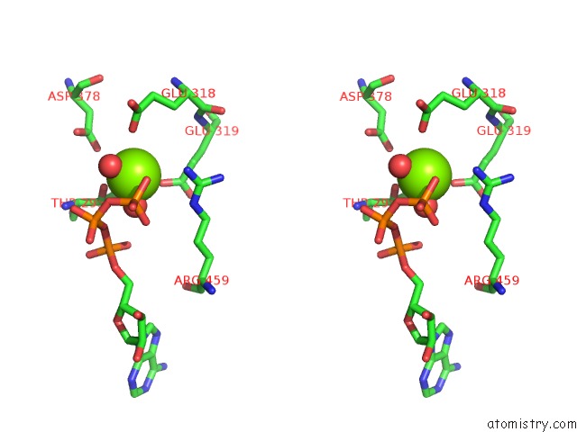
Magnesium binding site 4 out of 21 in 3s1a

Go back to Magnesium Binding Sites List in 3s1a
Magnesium binding site 4 out of 21 in the Crystal Structure of the Phosphorylation-Site Double Mutant S431E/T432E of the Kaic Circadian Clock Protein


Mono view


Stereo pair view

A full contact list of Magnesium with other atoms in the Mg binding site number 4 of Crystal Structure of the Phosphorylation-Site Double Mutant S431E/T432E of the Kaic Circadian Clock Protein within 5.0Å range:
probe atom residue distance (Å) B Occ
A:Mg526

b:58.1
occ:1.00
O1G F:ATP901 2.4 78.4 1.0
NZ A:LYS457 2.6 57.8 1.0
O2G F:ATP901 2.8 77.2 1.0
O A:GLU431 3.0 67.6 1.0
PG F:ATP901 3.1 77.8 1.0
CA A:GLU432 3.2 68.1 1.0
OG1 F:THR290 3.2 57.9 1.0
CG A:GLU432 3.5 66.1 1.0
O A:GLU432 3.8 67.0 1.0
C A:GLU431 3.8 66.0 1.0
NH2 A:ARG459 3.8 70.9 1.0
CE A:LYS457 3.9 61.9 1.0
CB A:GLU432 3.9 67.0 1.0
C A:GLU432 3.9 68.1 1.0
N A:GLU432 3.9 66.0 1.0
CB F:THR290 4.1 57.1 1.0
O3G F:ATP901 4.2 78.2 1.0
O3B F:ATP901 4.2 76.6 1.0
CA F:THR290 4.5 58.2 1.0
O A:THR434 4.6 70.7 1.0
CD A:LYS457 4.7 61.4 1.0
CZ A:ARG459 4.7 71.0 1.0
CD A:GLU432 4.8 66.3 1.0
OG1 F:THR415 4.9 68.3 1.0
OE2 F:GLU318 4.9 0.1 1.0
OE1 F:GLU318 5.0 0.1 1.0

Magnesium binding site 5 out of 21 in 3s1a

Go back to Magnesium Binding Sites List in 3s1a
Magnesium binding site 5 out of 21 in the Crystal Structure of the Phosphorylation-Site Double Mutant S431E/T432E of the Kaic Circadian Clock Protein


Mono view


Stereo pair view

A full contact list of Magnesium with other atoms in the Mg binding site number 5 of Crystal Structure of the Phosphorylation-Site Double Mutant S431E/T432E of the Kaic Circadian Clock Protein within 5.0Å range:
probe atom residue distance (Å) B Occ
B:Mg702

b:79.3
occ:1.00
O1G A:ATP903 2.3 77.0 1.0
CD1 B:PHE199 2.7 84.0 1.0
NZ B:LYS224 2.9 68.1 1.0
CA B:PHE199 3.1 85.6 1.0
CB B:PHE199 3.2 85.7 1.0
CG B:PHE199 3.2 85.0 1.0
OG A:SER48 3.4 41.6 1.0
PG A:ATP903 3.6 78.0 1.0
CE1 B:PHE199 3.6 84.0 1.0
O B:GLU198 3.7 86.8 1.0
CB A:SER48 3.7 41.4 1.0
N B:PHE199 3.8 86.4 1.0
CA A:SER48 3.8 42.8 1.0
CE B:LYS224 4.0 69.5 1.0
C B:GLU198 4.0 86.4 1.0
O3G A:ATP903 4.1 76.8 1.0
C B:PHE199 4.2 85.3 1.0
O3B A:ATP903 4.2 72.1 1.0
CD2 B:PHE199 4.3 85.0 1.0
O B:PHE199 4.3 85.2 1.0
NH2 B:ARG226 4.4 74.8 1.0
NZ A:LYS52 4.5 56.1 1.0
CD B:LYS224 4.7 69.8 1.0
CZ B:PHE199 4.7 84.7 1.0
C A:SER48 4.7 44.1 1.0
O2G A:ATP903 4.8 76.7 1.0
N A:SER48 4.8 42.3 1.0
N A:GLY49 4.8 43.7 1.0
CE2 B:PHE199 5.0 84.6 1.0

Magnesium binding site 6 out of 21 in 3s1a

Go back to Magnesium Binding Sites List in 3s1a
Magnesium binding site 6 out of 21 in the Crystal Structure of the Phosphorylation-Site Double Mutant S431E/T432E of the Kaic Circadian Clock Protein


Mono view


Stereo pair view

A full contact list of Magnesium with other atoms in the Mg binding site number 6 of Crystal Structure of the Phosphorylation-Site Double Mutant S431E/T432E of the Kaic Circadian Clock Protein within 5.0Å range:
probe atom residue distance (Å) B Occ
B:Mg801

b:45.4
occ:1.00
OG1 B:THR295 2.2 57.1 1.0
O B:HOH527 2.2 47.1 1.0
O2G B:ATP901 2.7 85.6 1.0
O B:HOH526 2.7 34.5 1.0
OE1 B:GLU318 2.7 92.3 1.0
O3B B:ATP901 2.9 84.1 1.0
O B:HOH530 2.9 37.0 1.0
PG B:ATP901 3.2 85.2 1.0
OD1 B:ASP378 3.5 70.8 1.0
CB B:THR295 3.5 58.2 1.0
O3G B:ATP901 3.6 87.7 1.0
OE2 B:GLU319 3.8 85.6 1.0
O3A B:ATP901 3.8 79.7 1.0
CD B:GLU318 3.9 92.2 1.0
PB B:ATP901 3.9 81.0 1.0
OD2 B:ASP378 4.2 70.9 1.0
CG B:ASP378 4.2 70.3 1.0
O1B B:ATP901 4.2 82.1 1.0
CG2 B:THR295 4.3 55.6 1.0
MG B:MG802 4.4 64.0 1.0
N B:THR295 4.5 58.3 1.0
CD B:GLU319 4.5 85.2 1.0
NH2 C:ARG459 4.5 63.0 1.0
CA B:THR295 4.5 58.4 1.0
O1G B:ATP901 4.6 86.6 1.0
OE2 B:GLU318 4.7 93.2 1.0
CG B:GLU319 4.8 85.0 1.0
CG B:GLU318 4.9 90.0 1.0
NZ B:LYS294 4.9 68.0 1.0
O2A B:ATP901 5.0 74.7 1.0

Magnesium binding site 7 out of 21 in 3s1a

Go back to Magnesium Binding Sites List in 3s1a
Magnesium binding site 7 out of 21 in the Crystal Structure of the Phosphorylation-Site Double Mutant S431E/T432E of the Kaic Circadian Clock Protein


Mono view


Stereo pair view

A full contact list of Magnesium with other atoms in the Mg binding site number 7 of Crystal Structure of the Phosphorylation-Site Double Mutant S431E/T432E of the Kaic Circadian Clock Protein within 5.0Å range:
probe atom residue distance (Å) B Occ
B:Mg802

b:64.0
occ:1.00
O1G B:ATP901 2.6 86.6 1.0
O2G B:ATP901 2.7 85.6 1.0
PG B:ATP901 2.9 85.2 1.0
O3B B:ATP901 3.2 84.1 1.0
O B:HOH530 3.3 37.0 1.0
OG1 B:THR415 3.3 65.9 1.0
CG C:GLU432 3.4 72.0 1.0
NZ B:LYS294 3.4 68.0 1.0
OG1 B:THR290 3.6 50.3 1.0
CA B:THR290 3.7 52.0 1.0
CG2 B:THR415 3.7 63.6 1.0
CB B:THR290 3.8 50.7 1.0
CA C:GLU432 3.9 66.8 1.0
CB B:THR415 4.1 64.9 1.0
NZ C:LYS457 4.1 47.3 1.0
CB C:GLU432 4.2 67.4 1.0
N B:GLY291 4.4 51.4 1.0
OE2 B:GLU318 4.4 93.2 1.0
O3G B:ATP901 4.4 87.7 1.0
MG B:MG801 4.4 45.4 1.0
OE1 B:GLU318 4.5 92.3 1.0
CD C:GLU432 4.6 75.0 1.0
PB B:ATP901 4.6 81.0 1.0
C B:THR290 4.6 52.4 1.0
CE B:LYS294 4.6 66.0 1.0
N B:THR290 4.7 53.2 1.0
O C:GLU431 4.7 69.3 1.0
NH2 C:ARG459 4.7 63.0 1.0
N C:GLU432 4.7 65.7 1.0
O C:GLU432 4.8 63.3 1.0
OE2 C:GLU432 4.8 76.3 1.0
C C:GLU432 4.8 65.1 1.0
CD B:GLU318 4.8 92.2 1.0
O2B B:ATP901 5.0 81.5 1.0

Magnesium binding site 8 out of 21 in 3s1a

Go back to Magnesium Binding Sites List in 3s1a
Magnesium binding site 8 out of 21 in the Crystal Structure of the Phosphorylation-Site Double Mutant S431E/T432E of the Kaic Circadian Clock Protein


Mono view


Stereo pair view

A full contact list of Magnesium with other atoms in the Mg binding site number 8 of Crystal Structure of the Phosphorylation-Site Double Mutant S431E/T432E of the Kaic Circadian Clock Protein within 5.0Å range:
probe atom residue distance (Å) B Occ
B:Mg701

b:58.5
occ:1.00
O2G B:ATP903 2.6 93.9 1.0
OG1 B:THR53 2.8 63.9 1.0
OD2 B:ASP145 3.0 92.0 1.0
O3B B:ATP903 3.2 92.5 1.0
PG B:ATP903 3.3 94.7 1.0
CG2 B:THR181 3.6 71.4 1.0
CE B:LYS52 3.7 59.7 1.0
OD1 B:ASP145 3.7 92.5 1.0
NZ B:LYS52 3.7 56.9 1.0
CG B:ASP145 3.7 91.4 1.0
O1G B:ATP903 3.7 94.6 1.0
CB B:LYS52 4.1 67.2 1.0
N B:THR53 4.1 67.2 1.0
CB B:THR53 4.2 64.9 1.0
PB B:ATP903 4.5 91.1 1.0
CA B:THR53 4.6 65.9 1.0
CD B:LYS52 4.6 61.6 1.0
CB B:SER146 4.6 92.2 1.0
O3G B:ATP903 4.7 95.8 1.0
CB B:THR181 4.7 71.3 1.0
OG1 B:THR181 4.8 69.9 1.0
O3A B:ATP903 4.8 88.6 1.0
O1B B:ATP903 4.8 90.3 1.0
OE1 B:GLU183 4.9 83.4 1.0
N B:SER146 4.9 90.4 1.0
CG B:LYS52 5.0 64.1 1.0

Magnesium binding site 9 out of 21 in 3s1a

Go back to Magnesium Binding Sites List in 3s1a
Magnesium binding site 9 out of 21 in the Crystal Structure of the Phosphorylation-Site Double Mutant S431E/T432E of the Kaic Circadian Clock Protein


Mono view


Stereo pair view

A full contact list of Magnesium with other atoms in the Mg binding site number 9 of Crystal Structure of the Phosphorylation-Site Double Mutant S431E/T432E of the Kaic Circadian Clock Protein within 5.0Å range:
probe atom residue distance (Å) B Occ
C:Mg802

b:41.9
occ:1.00
O1G C:ATP901 2.2 56.4 1.0
O2G C:ATP901 2.6 54.0 1.0
PG C:ATP901 2.7 56.3 1.0
OG1 C:THR415 3.2 41.8 1.0
O3B C:ATP901 3.2 53.7 1.0
CG D:GLU432 3.2 57.8 1.0
OG1 C:THR290 3.5 31.4 1.0
CB C:THR290 3.8 31.0 1.0
CA C:THR290 3.8 31.3 1.0
CG2 C:THR415 3.8 41.2 1.0
NZ C:LYS294 3.9 39.1 1.0
CA D:GLU432 4.0 55.1 1.0
CB C:THR415 4.0 41.5 1.0
O C:HOH527 4.0 25.2 1.0
NZ D:LYS457 4.1 29.1 1.0
O3G C:ATP901 4.2 56.9 1.0
CB D:GLU432 4.2 55.7 1.0
CD D:GLU432 4.3 59.6 1.0
OE2 D:GLU432 4.3 57.9 1.0
N C:GLY291 4.5 32.2 1.0
N D:GLU432 4.5 56.6 1.0
PB C:ATP901 4.6 49.5 1.0
OE1 C:GLU318 4.6 75.3 1.0
OE2 C:GLU318 4.6 77.8 1.0
C C:THR290 4.7 30.9 1.0
CE C:LYS294 4.8 40.0 1.0
NH2 D:ARG459 4.8 36.6 1.0
N C:THR290 4.9 30.5 1.0
C D:GLU431 5.0 57.1 1.0

Magnesium binding site 10 out of 21 in 3s1a

Go back to Magnesium Binding Sites List in 3s1a
Magnesium binding site 10 out of 21 in the Crystal Structure of the Phosphorylation-Site Double Mutant S431E/T432E of the Kaic Circadian Clock Protein


Mono view


Stereo pair view

A full contact list of Magnesium with other atoms in the Mg binding site number 10 of Crystal Structure of the Phosphorylation-Site Double Mutant S431E/T432E of the Kaic Circadian Clock Protein within 5.0Å range:
probe atom residue distance (Å) B Occ
C:Mg801

b:25.5
occ:1.00
O C:HOH526 1.9 30.0 1.0
O C:HOH527 1.9 25.2 1.0
OG1 C:THR295 2.3 41.4 1.0
OE2 C:GLU319 2.8 74.0 1.0
O2G C:ATP901 3.1 54.0 1.0
OE1 C:GLU318 3.2 75.3 1.0
CB C:THR295 3.4 45.7 1.0
O3B C:ATP901 3.4 53.7 1.0
PG C:ATP901 3.6 56.3 1.0
CG2 C:THR295 3.8 46.3 1.0
O3A C:ATP901 3.8 49.2 1.0
CD C:GLU319 3.9 72.5 1.0
O3G C:ATP901 3.9 56.9 1.0
NH2 D:ARG459 4.0 36.6 1.0
OD2 C:ASP378 4.0 57.3 1.0
PB C:ATP901 4.2 49.5 1.0
NE D:ARG459 4.3 37.8 1.0
CG C:GLU319 4.3 70.9 1.0
CD C:GLU318 4.4 75.7 1.0
CZ D:ARG459 4.4 37.6 1.0
O2A C:ATP901 4.6 50.9 1.0
O1B C:ATP901 4.6 53.5 1.0
CG C:ASP378 4.6 54.6 1.0
CA C:THR295 4.7 46.4 1.0
OD1 C:ASP378 4.7 52.3 1.0
PA C:ATP901 4.9 47.2 1.0
OE1 C:GLU319 4.9 73.6 1.0
N C:THR295 4.9 46.1 1.0

Reference:

R.Pattanayek, D.R.Williams, G.Rossi, S.Weigand, T.Mori, C.H.Johnson, P.L.Stewart, M.Egli. Combined Saxs/Em Based Models of the S. Elongatus Post-Translational Circadian Oscillator and Its Interactions with the Output His-Kinase Sasa. Plos One V. 6 23697 2011.
ISSN: ESSN 1932-6203
PubMed: 21887298
DOI: 10.1371/JOURNAL.PONE.0023697
Page generated: Mon Dec 14 08:48:45 2020

Last articles

Zn in 8WB0
Zn in 8WAX
Zn in 8WAU
Zn in 8WAZ
Zn in 8WAY
Zn in 8WAV
Zn in 8WAW
Zn in 8WAT
Zn in 8W7M
Zn in 8WD3
© Copyright 2008-2020 by atomistry.com
Home   |    Site Map   |    Copyright   |    Contact us   |    Privacy