Magnesium »
PDB 3s8c-3si7 »
3s9i »
Magnesium in PDB 3s9i: Crystal Structure of Mycobacterium Tuberculosis Malate Synthase in Complex with 2-4-Dioxo-4-Phenylbutanoic Acid Inhibitor
Enzymatic activity of Crystal Structure of Mycobacterium Tuberculosis Malate Synthase in Complex with 2-4-Dioxo-4-Phenylbutanoic Acid Inhibitor
All present enzymatic activity of Crystal Structure of Mycobacterium Tuberculosis Malate Synthase in Complex with 2-4-Dioxo-4-Phenylbutanoic Acid Inhibitor:
2.3.3.9;
2.3.3.9;
Protein crystallography data
The structure of Crystal Structure of Mycobacterium Tuberculosis Malate Synthase in Complex with 2-4-Dioxo-4-Phenylbutanoic Acid Inhibitor, PDB code: 3s9i
was solved by
I.V.Krieger,
Q.Sun,
J.C.Sacchettini,
Mycobacterium Tuberculosisstructural Proteomics Project (Xmtb),
with X-Ray Crystallography technique. A brief refinement statistics is given in the table below:
| Resolution Low / High (Å) | 44.99 / 1.90 |
| Space group | P 43 21 2 |
| Cell size a, b, c (Å), α, β, γ (°) | 79.331, 79.331, 225.945, 90.00, 90.00, 90.00 |
| R / Rfree (%) | 16.8 / 20.3 |
Magnesium Binding Sites:
The binding sites of Magnesium atom in the Crystal Structure of Mycobacterium Tuberculosis Malate Synthase in Complex with 2-4-Dioxo-4-Phenylbutanoic Acid Inhibitor
(pdb code 3s9i). This binding sites where shown within
5.0 Angstroms radius around Magnesium atom.
In total 3 binding sites of Magnesium where determined in the Crystal Structure of Mycobacterium Tuberculosis Malate Synthase in Complex with 2-4-Dioxo-4-Phenylbutanoic Acid Inhibitor, PDB code: 3s9i:
Jump to Magnesium binding site number: 1; 2; 3;
In total 3 binding sites of Magnesium where determined in the Crystal Structure of Mycobacterium Tuberculosis Malate Synthase in Complex with 2-4-Dioxo-4-Phenylbutanoic Acid Inhibitor, PDB code: 3s9i:
Jump to Magnesium binding site number: 1; 2; 3;
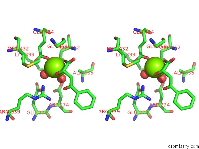
Magnesium binding site 1 out of 3 in 3s9i
Go back to
Magnesium binding site 1 out
of 3 in the Crystal Structure of Mycobacterium Tuberculosis Malate Synthase in Complex with 2-4-Dioxo-4-Phenylbutanoic Acid Inhibitor

Mono view
Stereo pair view
Mono view
Stereo pair view
A full contact list of Magnesium with other atoms in the Mg binding
site number 1 of Crystal Structure of Mycobacterium Tuberculosis Malate Synthase in Complex with 2-4-Dioxo-4-Phenylbutanoic Acid Inhibitor within 5.0Å range:
|
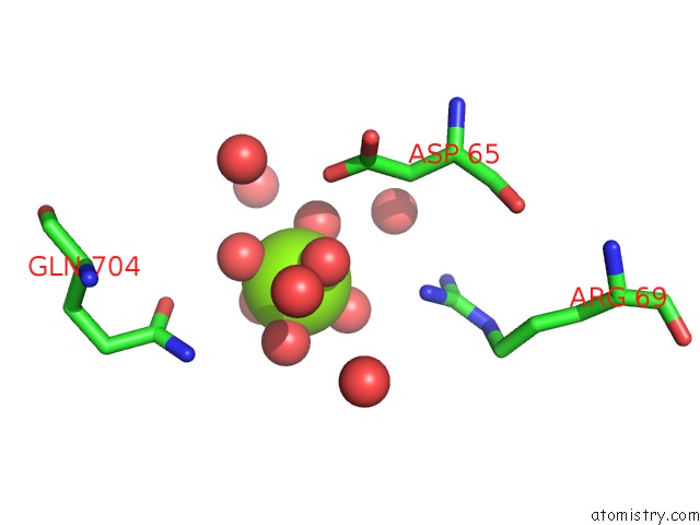
Magnesium binding site 2 out of 3 in 3s9i
Go back to
Magnesium binding site 2 out
of 3 in the Crystal Structure of Mycobacterium Tuberculosis Malate Synthase in Complex with 2-4-Dioxo-4-Phenylbutanoic Acid Inhibitor
Mono view
Stereo pair view
Mono view
Stereo pair view
A full contact list of Magnesium with other atoms in the Mg binding
site number 2 of Crystal Structure of Mycobacterium Tuberculosis Malate Synthase in Complex with 2-4-Dioxo-4-Phenylbutanoic Acid Inhibitor within 5.0Å range:
|
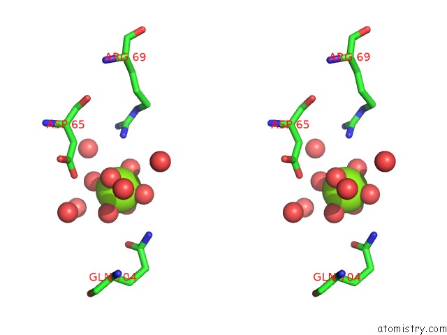
Magnesium binding site 3 out of 3 in 3s9i
Go back to
Magnesium binding site 3 out
of 3 in the Crystal Structure of Mycobacterium Tuberculosis Malate Synthase in Complex with 2-4-Dioxo-4-Phenylbutanoic Acid Inhibitor

Mono view

Stereo pair view
Mono view
Stereo pair view
A full contact list of Magnesium with other atoms in the Mg binding
site number 3 of Crystal Structure of Mycobacterium Tuberculosis Malate Synthase in Complex with 2-4-Dioxo-4-Phenylbutanoic Acid Inhibitor within 5.0Å range:
|
Reference:
I.V.Krieger,
J.S.Freundlich,
V.B.Gawandi,
J.P.Roberts,
V.B.Gawandi,
Q.Sun,
J.L.Owen,
M.T.Fraile,
S.I.Huss,
J.L.Lavandera,
T.R.Ioerger,
J.C.Sacchettini.
Structure-Guided Discovery of Phenyl-Diketo Acids As Potent Inhibitors of M. Tuberculosis Malate Synthase. Chem.Biol. V. 19 1556 2012.
ISSN: ISSN 1074-5521
PubMed: 23261599
DOI: 10.1016/J.CHEMBIOL.2012.09.018
Page generated: Thu Aug 15 10:49:06 2024
ISSN: ISSN 1074-5521
PubMed: 23261599
DOI: 10.1016/J.CHEMBIOL.2012.09.018
Last articles
Zn in 9J0NZn in 9J0O
Zn in 9J0P
Zn in 9FJX
Zn in 9EKB
Zn in 9C0F
Zn in 9CAH
Zn in 9CH0
Zn in 9CH3
Zn in 9CH1