Magnesium »
PDB 3s8c-3si7 »
3sf0 »
Magnesium in PDB 3sf0: Structure of Recombinant Haemophilus Influenzae E(P4) Acid Phosphatase Mutant D64N Complexed with 5'Amp
Enzymatic activity of Structure of Recombinant Haemophilus Influenzae E(P4) Acid Phosphatase Mutant D64N Complexed with 5'Amp
All present enzymatic activity of Structure of Recombinant Haemophilus Influenzae E(P4) Acid Phosphatase Mutant D64N Complexed with 5'Amp:
3.1.3.2;
3.1.3.2;
Protein crystallography data
The structure of Structure of Recombinant Haemophilus Influenzae E(P4) Acid Phosphatase Mutant D64N Complexed with 5'Amp, PDB code: 3sf0
was solved by
H.Singh,
with X-Ray Crystallography technique. A brief refinement statistics is given in the table below:
| Resolution Low / High (Å) | 36.12 / 1.35 |
| Space group | P 65 2 2 |
| Cell size a, b, c (Å), α, β, γ (°) | 97.897, 97.897, 107.009, 90.00, 90.00, 120.00 |
| R / Rfree (%) | 14.2 / 15.9 |
Magnesium Binding Sites:
The binding sites of Magnesium atom in the Structure of Recombinant Haemophilus Influenzae E(P4) Acid Phosphatase Mutant D64N Complexed with 5'Amp
(pdb code 3sf0). This binding sites where shown within
5.0 Angstroms radius around Magnesium atom.
In total only one binding site of Magnesium was determined in the Structure of Recombinant Haemophilus Influenzae E(P4) Acid Phosphatase Mutant D64N Complexed with 5'Amp, PDB code: 3sf0:
In total only one binding site of Magnesium was determined in the Structure of Recombinant Haemophilus Influenzae E(P4) Acid Phosphatase Mutant D64N Complexed with 5'Amp, PDB code: 3sf0:
Magnesium binding site 1 out of 1 in 3sf0
Go back to
Magnesium binding site 1 out
of 1 in the Structure of Recombinant Haemophilus Influenzae E(P4) Acid Phosphatase Mutant D64N Complexed with 5'Amp
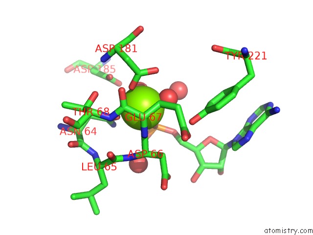
Mono view
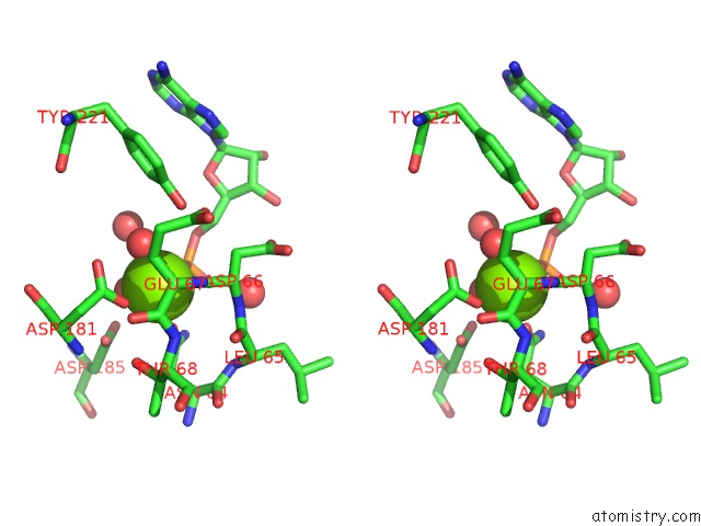
Stereo pair view
Mono view
Stereo pair view
A full contact list of Magnesium with other atoms in the Mg binding
site number 1 of Structure of Recombinant Haemophilus Influenzae E(P4) Acid Phosphatase Mutant D64N Complexed with 5'Amp within 5.0Å range:
|
Reference:
H.Singh,
T.J.Reilly,
J.J.Tanner.
Structural Basis of the Inhibition of Class C Acid Phosphatases By Adenosine 5'-Phosphorothioate. Febs J. V. 278 4374 2011.
ISSN: ISSN 1742-464X
PubMed: 21933344
DOI: 10.1111/J.1742-4658.2011.08360.X
Page generated: Thu Aug 15 10:55:13 2024
ISSN: ISSN 1742-464X
PubMed: 21933344
DOI: 10.1111/J.1742-4658.2011.08360.X
Last articles
Zn in 9J0NZn in 9J0O
Zn in 9J0P
Zn in 9FJX
Zn in 9EKB
Zn in 9C0F
Zn in 9CAH
Zn in 9CH0
Zn in 9CH3
Zn in 9CH1