Magnesium »
PDB 3s99-3si8 »
3sfu »
Magnesium in PDB 3sfu: Crystal Structure of Murine Norovirus Rna Dependent Rna Polymerase in Complex with Ribavirin
Protein crystallography data
The structure of Crystal Structure of Murine Norovirus Rna Dependent Rna Polymerase in Complex with Ribavirin, PDB code: 3sfu
was solved by
K.H.Kim,
I.Alam,
with X-Ray Crystallography technique. A brief refinement statistics is given in the table below:
| Resolution Low / High (Å) | 47.85 / 2.50 |
| Space group | C 1 2 1 |
| Cell size a, b, c (Å), α, β, γ (°) | 119.952, 196.084, 109.206, 90.00, 114.12, 90.00 |
| R / Rfree (%) | 18.1 / 22.9 |
Magnesium Binding Sites:
The binding sites of Magnesium atom in the Crystal Structure of Murine Norovirus Rna Dependent Rna Polymerase in Complex with Ribavirin
(pdb code 3sfu). This binding sites where shown within
5.0 Angstroms radius around Magnesium atom.
In total 3 binding sites of Magnesium where determined in the Crystal Structure of Murine Norovirus Rna Dependent Rna Polymerase in Complex with Ribavirin, PDB code: 3sfu:
Jump to Magnesium binding site number: 1; 2; 3;
In total 3 binding sites of Magnesium where determined in the Crystal Structure of Murine Norovirus Rna Dependent Rna Polymerase in Complex with Ribavirin, PDB code: 3sfu:
Jump to Magnesium binding site number: 1; 2; 3;
Magnesium binding site 1 out of 3 in 3sfu
Go back to
Magnesium binding site 1 out
of 3 in the Crystal Structure of Murine Norovirus Rna Dependent Rna Polymerase in Complex with Ribavirin
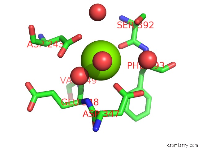
Mono view

Stereo pair view
Mono view
Stereo pair view
A full contact list of Magnesium with other atoms in the Mg binding
site number 1 of Crystal Structure of Murine Norovirus Rna Dependent Rna Polymerase in Complex with Ribavirin within 5.0Å range:
|
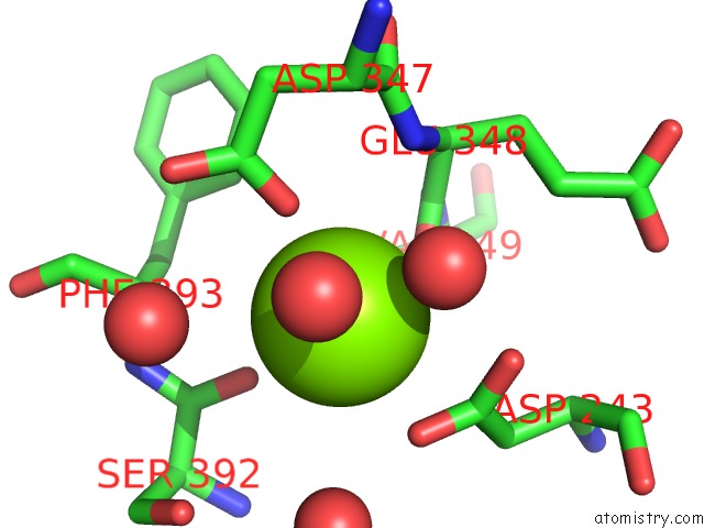
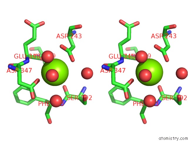
Magnesium binding site 2 out of 3 in 3sfu
Go back to
Magnesium binding site 2 out
of 3 in the Crystal Structure of Murine Norovirus Rna Dependent Rna Polymerase in Complex with Ribavirin

Mono view

Stereo pair view
Mono view
Stereo pair view
A full contact list of Magnesium with other atoms in the Mg binding
site number 2 of Crystal Structure of Murine Norovirus Rna Dependent Rna Polymerase in Complex with Ribavirin within 5.0Å range:
|
Magnesium binding site 3 out of 3 in 3sfu
Go back to
Magnesium binding site 3 out
of 3 in the Crystal Structure of Murine Norovirus Rna Dependent Rna Polymerase in Complex with Ribavirin
Mono view
Stereo pair view
Mono view
Stereo pair view
A full contact list of Magnesium with other atoms in the Mg binding
site number 3 of Crystal Structure of Murine Norovirus Rna Dependent Rna Polymerase in Complex with Ribavirin within 5.0Å range:
|
Reference:
I.Alam,
J.H.Lee,
K.J.Cho,
K.R.Han,
J.M.Yang,
M.S.Chung,
K.H.Kim.
Crystal Structures of Murine Norovirus-1 Rna-Dependent Rna Polymerase in Complex with 2-Thiouridine or Ribavirin. Virology V. 426 143 2012.
ISSN: ISSN 0042-6822
PubMed: 22341781
DOI: 10.1016/J.VIROL.2012.01.016
Page generated: Thu Aug 15 10:56:25 2024
ISSN: ISSN 0042-6822
PubMed: 22341781
DOI: 10.1016/J.VIROL.2012.01.016
Last articles
Fe in 2YXOFe in 2YRS
Fe in 2YXC
Fe in 2YNM
Fe in 2YVJ
Fe in 2YP1
Fe in 2YU2
Fe in 2YU1
Fe in 2YQB
Fe in 2YOO