Magnesium »
PDB 3t2d-3tar »
3t8o »
Magnesium in PDB 3t8o: Rhodopsin Kinase (GRK1) L166K Mutant at 2.5A Resolution
Enzymatic activity of Rhodopsin Kinase (GRK1) L166K Mutant at 2.5A Resolution
All present enzymatic activity of Rhodopsin Kinase (GRK1) L166K Mutant at 2.5A Resolution:
2.7.11.14;
2.7.11.14;
Protein crystallography data
The structure of Rhodopsin Kinase (GRK1) L166K Mutant at 2.5A Resolution, PDB code: 3t8o
was solved by
J.J.G.Tesmer,
P.Singh,
M.R.Nance,
with X-Ray Crystallography technique. A brief refinement statistics is given in the table below:
| Resolution Low / High (Å) | 29.60 / 2.50 |
| Space group | I 2 2 2 |
| Cell size a, b, c (Å), α, β, γ (°) | 55.541, 149.808, 190.879, 90.00, 90.00, 90.00 |
| R / Rfree (%) | 20.7 / 24.5 |
Other elements in 3t8o:
The structure of Rhodopsin Kinase (GRK1) L166K Mutant at 2.5A Resolution also contains other interesting chemical elements:
| Chlorine | (Cl) | 1 atom |
Magnesium Binding Sites:
The binding sites of Magnesium atom in the Rhodopsin Kinase (GRK1) L166K Mutant at 2.5A Resolution
(pdb code 3t8o). This binding sites where shown within
5.0 Angstroms radius around Magnesium atom.
In total only one binding site of Magnesium was determined in the Rhodopsin Kinase (GRK1) L166K Mutant at 2.5A Resolution, PDB code: 3t8o:
In total only one binding site of Magnesium was determined in the Rhodopsin Kinase (GRK1) L166K Mutant at 2.5A Resolution, PDB code: 3t8o:
Magnesium binding site 1 out of 1 in 3t8o
Go back to
Magnesium binding site 1 out
of 1 in the Rhodopsin Kinase (GRK1) L166K Mutant at 2.5A Resolution
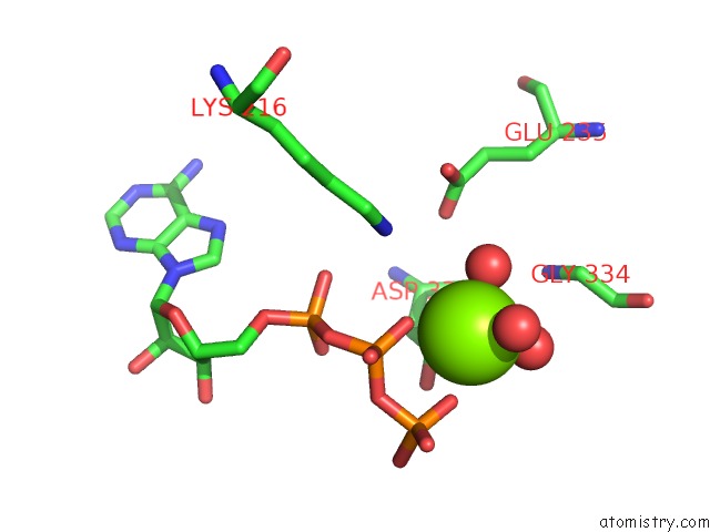
Mono view
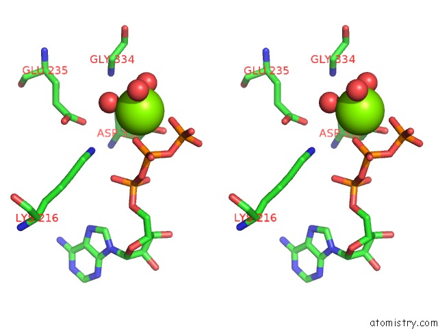
Stereo pair view
Mono view
Stereo pair view
A full contact list of Magnesium with other atoms in the Mg binding
site number 1 of Rhodopsin Kinase (GRK1) L166K Mutant at 2.5A Resolution within 5.0Å range:
|
Reference:
J.J.Tesmer,
M.R.Nance,
P.Singh,
H.Lee.
Structure of A Monomeric Variant of Rhodopsin Kinase at 2.5 A Resolution. Acta Crystallogr.,Sect.F V. 68 622 2012.
ISSN: ESSN 1744-3091
PubMed: 22684056
DOI: 10.1107/S1744309112017435
Page generated: Thu Aug 15 11:56:26 2024
ISSN: ESSN 1744-3091
PubMed: 22684056
DOI: 10.1107/S1744309112017435
Last articles
Fe in 2YXOFe in 2YRS
Fe in 2YXC
Fe in 2YNM
Fe in 2YVJ
Fe in 2YP1
Fe in 2YU2
Fe in 2YU1
Fe in 2YQB
Fe in 2YOO