Magnesium »
PDB 3tar-3tm0 »
3tba »
Magnesium in PDB 3tba: Structure of Yeast Ribonucleotide Reductase 1 Q288A with Dgtp and Adp
Enzymatic activity of Structure of Yeast Ribonucleotide Reductase 1 Q288A with Dgtp and Adp
All present enzymatic activity of Structure of Yeast Ribonucleotide Reductase 1 Q288A with Dgtp and Adp:
1.17.4.1;
1.17.4.1;
Protein crystallography data
The structure of Structure of Yeast Ribonucleotide Reductase 1 Q288A with Dgtp and Adp, PDB code: 3tba
was solved by
M.F.Ahmad,
P.S.Kaushal,
Q.Wan,
S.R.Wijeratna,
M.Huang,
C.Dealwis,
with X-Ray Crystallography technique. A brief refinement statistics is given in the table below:
| Resolution Low / High (Å) | 41.13 / 2.80 |
| Space group | P 21 21 2 |
| Cell size a, b, c (Å), α, β, γ (°) | 107.772, 118.086, 67.710, 90.00, 90.00, 90.00 |
| R / Rfree (%) | 19.6 / 26.4 |
Magnesium Binding Sites:
The binding sites of Magnesium atom in the Structure of Yeast Ribonucleotide Reductase 1 Q288A with Dgtp and Adp
(pdb code 3tba). This binding sites where shown within
5.0 Angstroms radius around Magnesium atom.
In total only one binding site of Magnesium was determined in the Structure of Yeast Ribonucleotide Reductase 1 Q288A with Dgtp and Adp, PDB code: 3tba:
In total only one binding site of Magnesium was determined in the Structure of Yeast Ribonucleotide Reductase 1 Q288A with Dgtp and Adp, PDB code: 3tba:
Magnesium binding site 1 out of 1 in 3tba
Go back to
Magnesium binding site 1 out
of 1 in the Structure of Yeast Ribonucleotide Reductase 1 Q288A with Dgtp and Adp
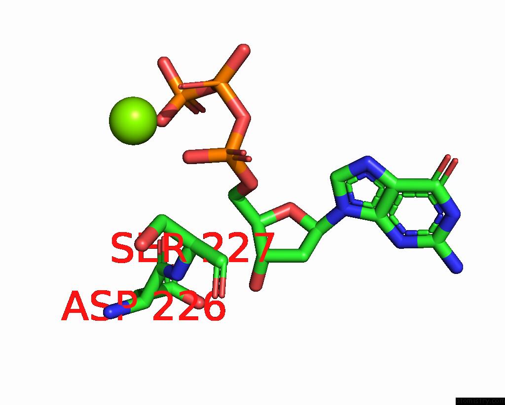
Mono view
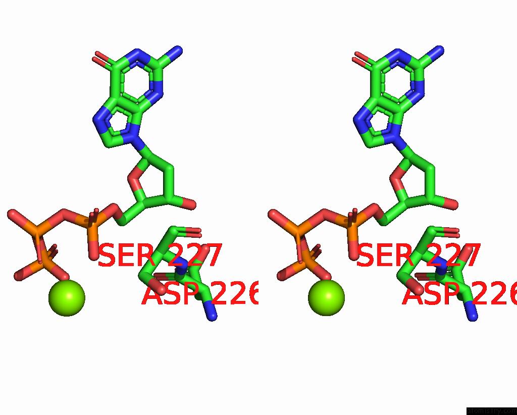
Stereo pair view
Mono view
Stereo pair view
A full contact list of Magnesium with other atoms in the Mg binding
site number 1 of Structure of Yeast Ribonucleotide Reductase 1 Q288A with Dgtp and Adp within 5.0Å range:
|
Reference:
M.F.Ahmad,
P.S.Kaushal,
Q.Wan,
S.R.Wijerathna,
X.An,
M.Huang,
C.G.Dealwis.
Role of Arginine 293 and Glutamine 288 in Communication Between Catalytic and Allosteric Sites in Yeast Ribonucleotide Reductase. J.Mol.Biol. V. 419 315 2012.
ISSN: ISSN 0022-2836
PubMed: 22465672
DOI: 10.1016/J.JMB.2012.03.014
Page generated: Thu Aug 15 12:02:51 2024
ISSN: ISSN 0022-2836
PubMed: 22465672
DOI: 10.1016/J.JMB.2012.03.014
Last articles
Zn in 9J0NZn in 9J0O
Zn in 9J0P
Zn in 9FJX
Zn in 9EKB
Zn in 9C0F
Zn in 9CAH
Zn in 9CH0
Zn in 9CH3
Zn in 9CH1