Magnesium »
PDB 3tar-3tm0 »
3thv »
Magnesium in PDB 3thv: Crystal Structure of Bacillus Dna Polymerase I Large Fragment Bound to Dna and Ddatp-Dt in Closed Conformation
Enzymatic activity of Crystal Structure of Bacillus Dna Polymerase I Large Fragment Bound to Dna and Ddatp-Dt in Closed Conformation
All present enzymatic activity of Crystal Structure of Bacillus Dna Polymerase I Large Fragment Bound to Dna and Ddatp-Dt in Closed Conformation:
2.7.7.7;
2.7.7.7;
Protein crystallography data
The structure of Crystal Structure of Bacillus Dna Polymerase I Large Fragment Bound to Dna and Ddatp-Dt in Closed Conformation, PDB code: 3thv
was solved by
W.Wang,
L.S.Beese,
with X-Ray Crystallography technique. A brief refinement statistics is given in the table below:
| Resolution Low / High (Å) | 88.41 / 1.61 |
| Space group | P 21 21 21 |
| Cell size a, b, c (Å), α, β, γ (°) | 93.810, 109.270, 150.430, 90.00, 90.00, 90.00 |
| R / Rfree (%) | 19.3 / 22.4 |
Magnesium Binding Sites:
The binding sites of Magnesium atom in the Crystal Structure of Bacillus Dna Polymerase I Large Fragment Bound to Dna and Ddatp-Dt in Closed Conformation
(pdb code 3thv). This binding sites where shown within
5.0 Angstroms radius around Magnesium atom.
In total 2 binding sites of Magnesium where determined in the Crystal Structure of Bacillus Dna Polymerase I Large Fragment Bound to Dna and Ddatp-Dt in Closed Conformation, PDB code: 3thv:
Jump to Magnesium binding site number: 1; 2;
In total 2 binding sites of Magnesium where determined in the Crystal Structure of Bacillus Dna Polymerase I Large Fragment Bound to Dna and Ddatp-Dt in Closed Conformation, PDB code: 3thv:
Jump to Magnesium binding site number: 1; 2;
Magnesium binding site 1 out of 2 in 3thv
Go back to
Magnesium binding site 1 out
of 2 in the Crystal Structure of Bacillus Dna Polymerase I Large Fragment Bound to Dna and Ddatp-Dt in Closed Conformation
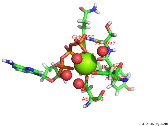
Mono view
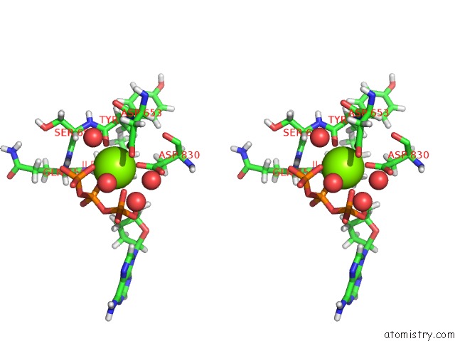
Stereo pair view
Mono view
Stereo pair view
A full contact list of Magnesium with other atoms in the Mg binding
site number 1 of Crystal Structure of Bacillus Dna Polymerase I Large Fragment Bound to Dna and Ddatp-Dt in Closed Conformation within 5.0Å range:
|
Magnesium binding site 2 out of 2 in 3thv
Go back to
Magnesium binding site 2 out
of 2 in the Crystal Structure of Bacillus Dna Polymerase I Large Fragment Bound to Dna and Ddatp-Dt in Closed Conformation
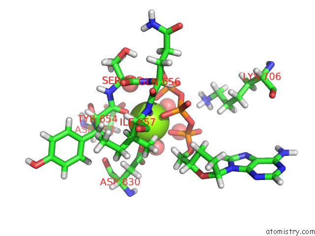
Mono view
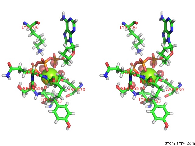
Stereo pair view
Mono view
Stereo pair view
A full contact list of Magnesium with other atoms in the Mg binding
site number 2 of Crystal Structure of Bacillus Dna Polymerase I Large Fragment Bound to Dna and Ddatp-Dt in Closed Conformation within 5.0Å range:
|
Reference:
W.Wang,
H.W.Hellinga,
L.S.Beese.
Structural Evidence For the Rare Tautomer Hypothesis of Spontaneous Mutagenesis. Proc.Natl.Acad.Sci.Usa V. 108 17644 2011.
ISSN: ISSN 0027-8424
PubMed: 22006298
DOI: 10.1073/PNAS.1114496108
Page generated: Thu Aug 15 12:07:48 2024
ISSN: ISSN 0027-8424
PubMed: 22006298
DOI: 10.1073/PNAS.1114496108
Last articles
Zn in 9J0NZn in 9J0O
Zn in 9J0P
Zn in 9FJX
Zn in 9EKB
Zn in 9C0F
Zn in 9CAH
Zn in 9CH0
Zn in 9CH3
Zn in 9CH1