Magnesium »
PDB 3tav-3tnf »
3tin »
Magnesium in PDB 3tin: Tubulin Tyrosine Ligase
Protein crystallography data
The structure of Tubulin Tyrosine Ligase, PDB code: 3tin
was solved by
A.Roll-Mecak,
A.Szyk,
A.Deaconescu,
G.Piszczek,
with X-Ray Crystallography technique. A brief refinement statistics is given in the table below:
| Resolution Low / High (Å) | 29.24 / 2.90 |
| Space group | C 1 2 1 |
| Cell size a, b, c (Å), α, β, γ (°) | 116.970, 75.720, 44.230, 90.00, 90.83, 90.00 |
| R / Rfree (%) | 23.2 / 31.3 |
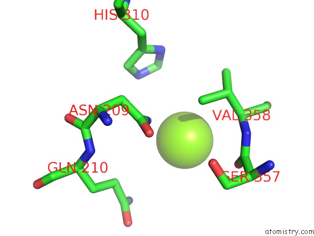
Magnesium Binding Sites:
The binding sites of Magnesium atom in the Tubulin Tyrosine Ligase
(pdb code 3tin). This binding sites where shown within
5.0 Angstroms radius around Magnesium atom.
In total 3 binding sites of Magnesium where determined in the Tubulin Tyrosine Ligase, PDB code: 3tin:
Jump to Magnesium binding site number: 1; 2; 3;
In total 3 binding sites of Magnesium where determined in the Tubulin Tyrosine Ligase, PDB code: 3tin:
Jump to Magnesium binding site number: 1; 2; 3;
Magnesium binding site 1 out of 3 in 3tin
Go back to
Magnesium binding site 1 out
of 3 in the Tubulin Tyrosine Ligase
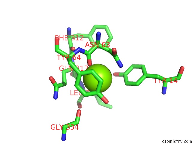
Mono view
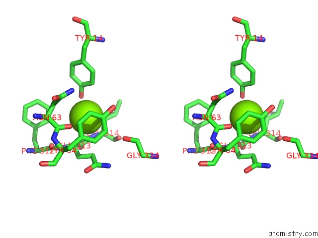
Stereo pair view
Mono view
Stereo pair view
A full contact list of Magnesium with other atoms in the Mg binding
site number 1 of Tubulin Tyrosine Ligase within 5.0Å range:
|
Magnesium binding site 2 out of 3 in 3tin
Go back to
Magnesium binding site 2 out
of 3 in the Tubulin Tyrosine Ligase
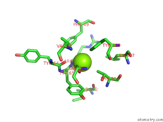
Mono view
Stereo pair view
Mono view
Stereo pair view
A full contact list of Magnesium with other atoms in the Mg binding
site number 2 of Tubulin Tyrosine Ligase within 5.0Å range:
|
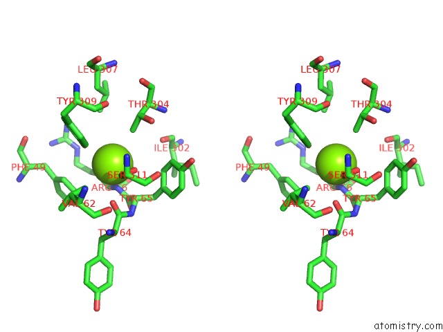
Magnesium binding site 3 out of 3 in 3tin
Go back to
Magnesium binding site 3 out
of 3 in the Tubulin Tyrosine Ligase
Mono view

Stereo pair view
Mono view
Stereo pair view
A full contact list of Magnesium with other atoms in the Mg binding
site number 3 of Tubulin Tyrosine Ligase within 5.0Å range:
|
Reference:
A.Szyk,
A.M.Deaconescu,
G.Piszczek,
A.Roll-Mecak.
Tubulin Tyrosine Ligase Structure Reveals Adaptation of An Ancient Fold to Bind and Modify Tubulin. Nat.Struct.Mol.Biol. V. 18 1250 2011.
ISSN: ISSN 1545-9993
PubMed: 22020298
DOI: 10.1038/NSMB.2148
Page generated: Thu Aug 15 12:08:46 2024
ISSN: ISSN 1545-9993
PubMed: 22020298
DOI: 10.1038/NSMB.2148
Last articles
Cl in 7SUHCl in 7SQE
Cl in 7STV
Cl in 7STT
Cl in 7STQ
Cl in 7SSF
Cl in 7SSB
Cl in 7SS8
Cl in 7SR6
Cl in 7SS7