Magnesium »
PDB 3tar-3tm0 »
3tlm »
Magnesium in PDB 3tlm: Crystal Structure of Endoplasmic Reticulum CA2+-Atpase (Serca) From Bovine Muscle
Enzymatic activity of Crystal Structure of Endoplasmic Reticulum CA2+-Atpase (Serca) From Bovine Muscle
All present enzymatic activity of Crystal Structure of Endoplasmic Reticulum CA2+-Atpase (Serca) From Bovine Muscle:
3.6.3.8;
3.6.3.8;
Protein crystallography data
The structure of Crystal Structure of Endoplasmic Reticulum CA2+-Atpase (Serca) From Bovine Muscle, PDB code: 3tlm
was solved by
R.Sacchetto,
I.Bertipaglia,
S.Giannetti,
L.Cendron,
F.Mascarello,
E.Damiani,
E.Carafoli,
G.Zanotti,
with X-Ray Crystallography technique. A brief refinement statistics is given in the table below:
| Resolution Low / High (Å) | 41.99 / 2.95 |
| Space group | C 1 2 1 |
| Cell size a, b, c (Å), α, β, γ (°) | 156.580, 75.260, 151.800, 90.00, 108.30, 90.00 |
| R / Rfree (%) | 20.5 / 27.7 |
Other elements in 3tlm:
The structure of Crystal Structure of Endoplasmic Reticulum CA2+-Atpase (Serca) From Bovine Muscle also contains other interesting chemical elements:
| Potassium | (K) | 1 atom |
| Calcium | (Ca) | 2 atoms |
Magnesium Binding Sites:
The binding sites of Magnesium atom in the Crystal Structure of Endoplasmic Reticulum CA2+-Atpase (Serca) From Bovine Muscle
(pdb code 3tlm). This binding sites where shown within
5.0 Angstroms radius around Magnesium atom.
In total only one binding site of Magnesium was determined in the Crystal Structure of Endoplasmic Reticulum CA2+-Atpase (Serca) From Bovine Muscle, PDB code: 3tlm:
In total only one binding site of Magnesium was determined in the Crystal Structure of Endoplasmic Reticulum CA2+-Atpase (Serca) From Bovine Muscle, PDB code: 3tlm:
Magnesium binding site 1 out of 1 in 3tlm
Go back to
Magnesium binding site 1 out
of 1 in the Crystal Structure of Endoplasmic Reticulum CA2+-Atpase (Serca) From Bovine Muscle
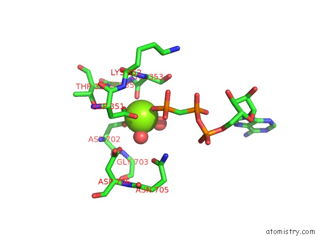
Mono view
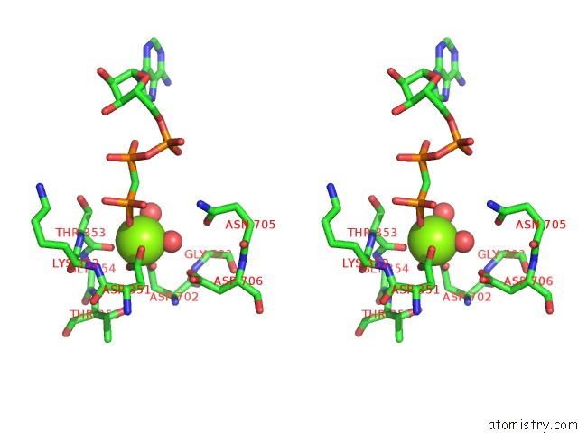
Stereo pair view
Mono view
Stereo pair view
A full contact list of Magnesium with other atoms in the Mg binding
site number 1 of Crystal Structure of Endoplasmic Reticulum CA2+-Atpase (Serca) From Bovine Muscle within 5.0Å range:
|
Reference:
R.Sacchetto,
I.Bertipaglia,
S.Giannetti,
L.Cendron,
F.Mascarello,
E.Damiani,
E.Carafoli,
G.Zanotti.
Crystal Structure of Sarcoplasmic Reticulum Ca(2+)-Atpase (Serca) From Bovine Muscle. J.Struct.Biol. V. 178 38 2012.
ISSN: ISSN 1047-8477
PubMed: 22387132
DOI: 10.1016/J.JSB.2012.02.008
Page generated: Thu Aug 15 12:09:57 2024
ISSN: ISSN 1047-8477
PubMed: 22387132
DOI: 10.1016/J.JSB.2012.02.008
Last articles
Zn in 9J0NZn in 9J0O
Zn in 9J0P
Zn in 9FJX
Zn in 9EKB
Zn in 9C0F
Zn in 9CAH
Zn in 9CH0
Zn in 9CH3
Zn in 9CH1