Magnesium »
PDB 3tnq-3twp »
3tte »
Magnesium in PDB 3tte: Crystal Structure of Enolase BRADO_4202 (Target Efi-501651) From Bradyrhizobium Complexed with Magnesium and Mandelic Acid
Enzymatic activity of Crystal Structure of Enolase BRADO_4202 (Target Efi-501651) From Bradyrhizobium Complexed with Magnesium and Mandelic Acid
All present enzymatic activity of Crystal Structure of Enolase BRADO_4202 (Target Efi-501651) From Bradyrhizobium Complexed with Magnesium and Mandelic Acid:
5.1.2.2; 5.5.1.1;
5.1.2.2; 5.5.1.1;
Protein crystallography data
The structure of Crystal Structure of Enolase BRADO_4202 (Target Efi-501651) From Bradyrhizobium Complexed with Magnesium and Mandelic Acid, PDB code: 3tte
was solved by
Y.Patskovsky,
J.Kim,
R.Toro,
R.Bhosle,
B.Hillerich,
R.D.Seidel,
E.Washington,
A.Scott Glenn,
S.Chowdhury,
B.Evans,
J.Hammond,
W.D.Zencheck,
H.J.Imker,
J.A.Gerlt,
S.C.Almo,
Enzyme Function Initiative(Efi),
with X-Ray Crystallography technique. A brief refinement statistics is given in the table below:
| Resolution Low / High (Å) | 50.00 / 2.00 |
| Space group | I 2 2 2 |
| Cell size a, b, c (Å), α, β, γ (°) | 109.949, 118.867, 130.571, 90.00, 90.00, 90.00 |
| R / Rfree (%) | 20.4 / 26.3 |
Magnesium Binding Sites:
The binding sites of Magnesium atom in the Crystal Structure of Enolase BRADO_4202 (Target Efi-501651) From Bradyrhizobium Complexed with Magnesium and Mandelic Acid
(pdb code 3tte). This binding sites where shown within
5.0 Angstroms radius around Magnesium atom.
In total 2 binding sites of Magnesium where determined in the Crystal Structure of Enolase BRADO_4202 (Target Efi-501651) From Bradyrhizobium Complexed with Magnesium and Mandelic Acid, PDB code: 3tte:
Jump to Magnesium binding site number: 1; 2;
In total 2 binding sites of Magnesium where determined in the Crystal Structure of Enolase BRADO_4202 (Target Efi-501651) From Bradyrhizobium Complexed with Magnesium and Mandelic Acid, PDB code: 3tte:
Jump to Magnesium binding site number: 1; 2;
Magnesium binding site 1 out of 2 in 3tte
Go back to
Magnesium binding site 1 out
of 2 in the Crystal Structure of Enolase BRADO_4202 (Target Efi-501651) From Bradyrhizobium Complexed with Magnesium and Mandelic Acid
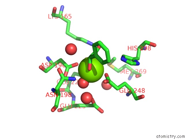
Mono view
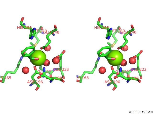
Stereo pair view
Mono view
Stereo pair view
A full contact list of Magnesium with other atoms in the Mg binding
site number 1 of Crystal Structure of Enolase BRADO_4202 (Target Efi-501651) From Bradyrhizobium Complexed with Magnesium and Mandelic Acid within 5.0Å range:
|
Magnesium binding site 2 out of 2 in 3tte
Go back to
Magnesium binding site 2 out
of 2 in the Crystal Structure of Enolase BRADO_4202 (Target Efi-501651) From Bradyrhizobium Complexed with Magnesium and Mandelic Acid
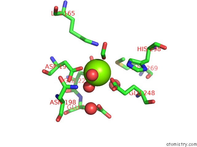
Mono view
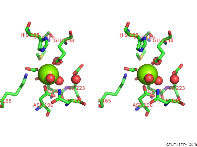
Stereo pair view
Mono view
Stereo pair view
A full contact list of Magnesium with other atoms in the Mg binding
site number 2 of Crystal Structure of Enolase BRADO_4202 (Target Efi-501651) From Bradyrhizobium Complexed with Magnesium and Mandelic Acid within 5.0Å range:
|
Reference:
Y.Patskovsky,
J.Kim,
R.Toro,
R.Bhosle,
B.Hillerich,
R.D.Seidel,
E.Washington,
A.Scott Glenn,
S.Chowdhury,
B.Evans,
J.Hammond,
W.D.Zencheck,
H.J.Imker,
J.A.Gerlt,
S.C.Almo.
Crystal Structure of Mandelate Racemase From Bradyrhizobium Sp. ORS278 To Be Published.
Page generated: Mon Aug 11 03:59:46 2025
Last articles
Mg in 4DV3Mg in 4DV2
Mg in 4DV0
Mg in 4DV1
Mg in 4DUZ
Mg in 4DUY
Mg in 4DR7
Mg in 4DR6
Mg in 4DR5
Mg in 4DUX