Magnesium »
PDB 3tnq-3twp »
3tvb »
Magnesium in PDB 3tvb: A Highly Symmetric Dna G-4 Quadruplex/Drug Complex
Protein crystallography data
The structure of A Highly Symmetric Dna G-4 Quadruplex/Drug Complex, PDB code: 3tvb
was solved by
G.R.Clark,
P.D.Pytel,
C.J.Squire,
with X-Ray Crystallography technique. A brief refinement statistics is given in the table below:
| Resolution Low / High (Å) | 10.00 / 1.08 |
| Space group | I 4 |
| Cell size a, b, c (Å), α, β, γ (°) | 40.209, 40.209, 49.831, 90.00, 90.00, 90.00 |
| R / Rfree (%) | 16.2 / 20.2 |
Other elements in 3tvb:
The structure of A Highly Symmetric Dna G-4 Quadruplex/Drug Complex also contains other interesting chemical elements:
| Sodium | (Na) | 8 atoms |
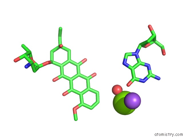
Magnesium Binding Sites:
The binding sites of Magnesium atom in the A Highly Symmetric Dna G-4 Quadruplex/Drug Complex
(pdb code 3tvb). This binding sites where shown within
5.0 Angstroms radius around Magnesium atom.
In total 2 binding sites of Magnesium where determined in the A Highly Symmetric Dna G-4 Quadruplex/Drug Complex, PDB code: 3tvb:
Jump to Magnesium binding site number: 1; 2;
In total 2 binding sites of Magnesium where determined in the A Highly Symmetric Dna G-4 Quadruplex/Drug Complex, PDB code: 3tvb:
Jump to Magnesium binding site number: 1; 2;
Magnesium binding site 1 out of 2 in 3tvb
Go back to
Magnesium binding site 1 out
of 2 in the A Highly Symmetric Dna G-4 Quadruplex/Drug Complex
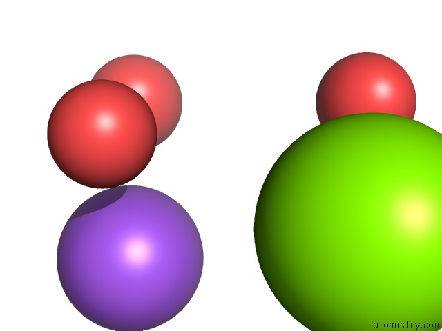
Mono view
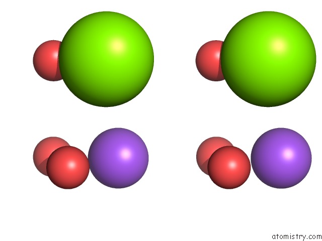
Stereo pair view
Mono view
Stereo pair view
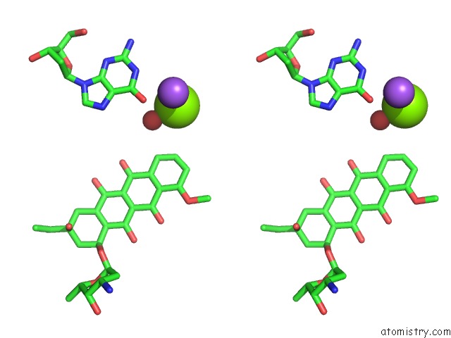
A full contact list of Magnesium with other atoms in the Mg binding
site number 1 of A Highly Symmetric Dna G-4 Quadruplex/Drug Complex within 5.0Å range:
|
Magnesium binding site 2 out of 2 in 3tvb
Go back to
Magnesium binding site 2 out
of 2 in the A Highly Symmetric Dna G-4 Quadruplex/Drug Complex
Mono view
Stereo pair view
Mono view
Stereo pair view
A full contact list of Magnesium with other atoms in the Mg binding
site number 2 of A Highly Symmetric Dna G-4 Quadruplex/Drug Complex within 5.0Å range:
|
Reference:
G.R.Clark,
P.D.Pytel,
C.J.Squire.
The High-Resolution Crystal Structure of A Parallel Intermolecular Dna G-4 Quadruplex/Drug Complex Employing Syn Glycosyl Linkages. Nucleic Acids Res. V. 40 5731 2012.
ISSN: ISSN 0305-1048
PubMed: 22373921
DOI: 10.1093/NAR/GKS193
Page generated: Mon Aug 11 04:00:12 2025
ISSN: ISSN 0305-1048
PubMed: 22373921
DOI: 10.1093/NAR/GKS193
Last articles
Mg in 4B13Mg in 4B1U
Mg in 4AZC
Mg in 4B12
Mg in 4B11
Mg in 4B10
Mg in 4B0T
Mg in 4B0S
Mg in 4B0H
Mg in 4AZ5