Magnesium »
PDB 3tnq-3twp »
3tvd »
Magnesium in PDB 3tvd: Crystal Structure of Mouse Rhoa-Gtp Complex
Protein crystallography data
The structure of Crystal Structure of Mouse Rhoa-Gtp Complex, PDB code: 3tvd
was solved by
K.Swaminathan,
K.Pal,
C.Jobichen,
with X-Ray Crystallography technique. A brief refinement statistics is given in the table below:
| Resolution Low / High (Å) | 15.00 / 2.99 |
| Space group | C 2 2 2 |
| Cell size a, b, c (Å), α, β, γ (°) | 80.813, 122.652, 91.276, 90.00, 90.00, 90.00 |
| R / Rfree (%) | 22.3 / 28.2 |
Magnesium Binding Sites:
The binding sites of Magnesium atom in the Crystal Structure of Mouse Rhoa-Gtp Complex
(pdb code 3tvd). This binding sites where shown within
5.0 Angstroms radius around Magnesium atom.
In total 2 binding sites of Magnesium where determined in the Crystal Structure of Mouse Rhoa-Gtp Complex, PDB code: 3tvd:
Jump to Magnesium binding site number: 1; 2;
In total 2 binding sites of Magnesium where determined in the Crystal Structure of Mouse Rhoa-Gtp Complex, PDB code: 3tvd:
Jump to Magnesium binding site number: 1; 2;
Magnesium binding site 1 out of 2 in 3tvd
Go back to
Magnesium binding site 1 out
of 2 in the Crystal Structure of Mouse Rhoa-Gtp Complex
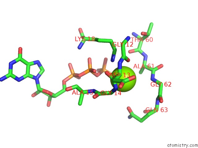
Mono view
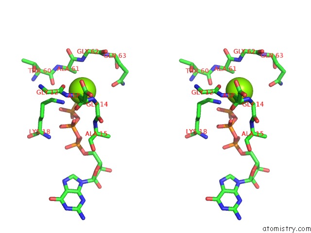
Stereo pair view
Mono view
Stereo pair view
A full contact list of Magnesium with other atoms in the Mg binding
site number 1 of Crystal Structure of Mouse Rhoa-Gtp Complex within 5.0Å range:
|
Magnesium binding site 2 out of 2 in 3tvd
Go back to
Magnesium binding site 2 out
of 2 in the Crystal Structure of Mouse Rhoa-Gtp Complex
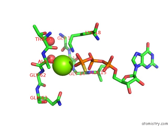
Mono view
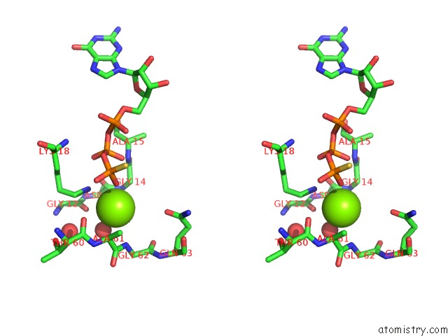
Stereo pair view
Mono view
Stereo pair view
A full contact list of Magnesium with other atoms in the Mg binding
site number 2 of Crystal Structure of Mouse Rhoa-Gtp Complex within 5.0Å range:
|
Reference:
C.Jobichen,
K.Pal,
K.Swaminathan.
Crystal Structure of Mouse Rhoa:Gtpgammas Complex in A Centered Lattice. J.Struct.Funct.Genom. V. 13 241 2012.
ISSN: ISSN 1345-711X
PubMed: 23001747
DOI: 10.1007/S10969-012-9143-5
Page generated: Mon Aug 11 04:00:28 2025
ISSN: ISSN 1345-711X
PubMed: 23001747
DOI: 10.1007/S10969-012-9143-5
Last articles
Mg in 5BTLMg in 5BTM
Mg in 5BTI
Mg in 5BTG
Mg in 5BTF
Mg in 5BTD
Mg in 5BTC
Mg in 5BTA
Mg in 5BON
Mg in 5BSU