Magnesium »
PDB 3twp-3u89 »
3txz »
Magnesium in PDB 3txz: OYE1-W116Q Complexed with R-Carvone
Enzymatic activity of OYE1-W116Q Complexed with R-Carvone
All present enzymatic activity of OYE1-W116Q Complexed with R-Carvone:
1.6.99.1;
1.6.99.1;
Protein crystallography data
The structure of OYE1-W116Q Complexed with R-Carvone, PDB code: 3txz
was solved by
B.Sullivan,
Y.A.Pompeu,
J.D.Stewart,
with X-Ray Crystallography technique. A brief refinement statistics is given in the table below:
| Resolution Low / High (Å) | 45.00 / 1.70 |
| Space group | P 43 21 2 |
| Cell size a, b, c (Å), α, β, γ (°) | 141.362, 141.362, 42.687, 90.00, 90.00, 90.00 |
| R / Rfree (%) | 16.1 / 18.6 |
Magnesium Binding Sites:
The binding sites of Magnesium atom in the OYE1-W116Q Complexed with R-Carvone
(pdb code 3txz). This binding sites where shown within
5.0 Angstroms radius around Magnesium atom.
In total only one binding site of Magnesium was determined in the OYE1-W116Q Complexed with R-Carvone, PDB code: 3txz:
In total only one binding site of Magnesium was determined in the OYE1-W116Q Complexed with R-Carvone, PDB code: 3txz:
Magnesium binding site 1 out of 1 in 3txz
Go back to
Magnesium binding site 1 out
of 1 in the OYE1-W116Q Complexed with R-Carvone
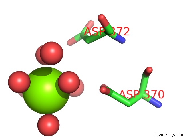
Mono view
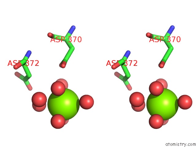
Stereo pair view
Mono view
Stereo pair view
A full contact list of Magnesium with other atoms in the Mg binding
site number 1 of OYE1-W116Q Complexed with R-Carvone within 5.0Å range:
|
Reference:
Y.A.Pompeu,
B.Sullivan,
J.D.Stewart.
X‑Ray Crystallography Reveals How Subtle Changes Control the Orientation of Substrate Binding in An Alkene Reductase Acs Catalysis V. 3 2376 2013.
ISSN: ESSN 2155-5435
DOI: 10.1021/CS400622E
Page generated: Thu Aug 15 12:16:58 2024
ISSN: ESSN 2155-5435
DOI: 10.1021/CS400622E
Last articles
Zn in 9JYWZn in 9IR4
Zn in 9IR3
Zn in 9GMX
Zn in 9GMW
Zn in 9JEJ
Zn in 9ERF
Zn in 9ERE
Zn in 9EGV
Zn in 9EGW