Magnesium »
PDB 3u8x-3ump »
3u9d »
Magnesium in PDB 3u9d: Crystal Structure of A Chimera Containing the N-Terminal Domain (Residues 8-24) of Drosophila Ciboulot and the C-Terminal Domain (Residues 13-44) of Bovine Thymosin-BETA4, Bound to G-Actin-Atp
Protein crystallography data
The structure of Crystal Structure of A Chimera Containing the N-Terminal Domain (Residues 8-24) of Drosophila Ciboulot and the C-Terminal Domain (Residues 13-44) of Bovine Thymosin-BETA4, Bound to G-Actin-Atp, PDB code: 3u9d
was solved by
L.Renault,
C.Husson,
M.F.Carlier,
D.Didry,
with X-Ray Crystallography technique. A brief refinement statistics is given in the table below:
| Resolution Low / High (Å) | 19.64 / 2.50 |
| Space group | P 1 21 1 |
| Cell size a, b, c (Å), α, β, γ (°) | 45.698, 75.738, 128.518, 90.00, 90.03, 90.00 |
| R / Rfree (%) | 20.4 / 24 |
Magnesium Binding Sites:
The binding sites of Magnesium atom in the Crystal Structure of A Chimera Containing the N-Terminal Domain (Residues 8-24) of Drosophila Ciboulot and the C-Terminal Domain (Residues 13-44) of Bovine Thymosin-BETA4, Bound to G-Actin-Atp
(pdb code 3u9d). This binding sites where shown within
5.0 Angstroms radius around Magnesium atom.
In total 2 binding sites of Magnesium where determined in the Crystal Structure of A Chimera Containing the N-Terminal Domain (Residues 8-24) of Drosophila Ciboulot and the C-Terminal Domain (Residues 13-44) of Bovine Thymosin-BETA4, Bound to G-Actin-Atp, PDB code: 3u9d:
Jump to Magnesium binding site number: 1; 2;
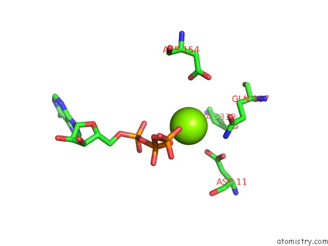
Magnesium binding site 1 out of 2 in 3u9d
Go back to
Magnesium binding site 1 out
of 2 in the Crystal Structure of A Chimera Containing the N-Terminal Domain (Residues 8-24) of Drosophila Ciboulot and the C-Terminal Domain (Residues 13-44) of Bovine Thymosin-BETA4, Bound to G-Actin-Atp
Mono view
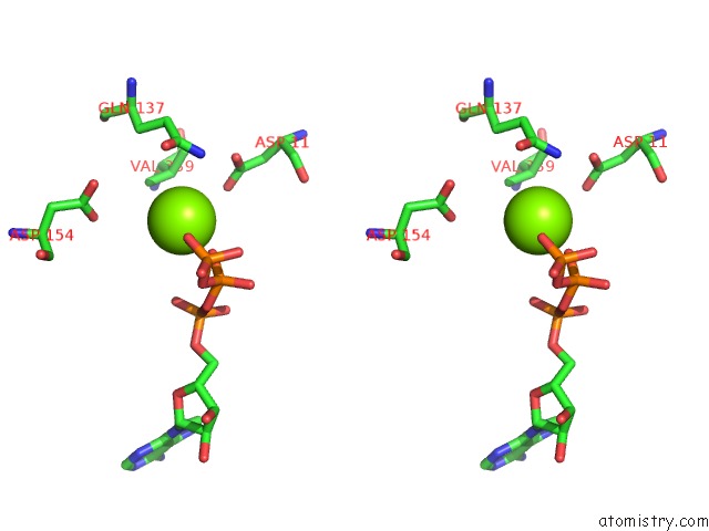
Stereo pair view
Mono view
Stereo pair view
A full contact list of Magnesium with other atoms in the Mg binding
site number 1 of Crystal Structure of A Chimera Containing the N-Terminal Domain (Residues 8-24) of Drosophila Ciboulot and the C-Terminal Domain (Residues 13-44) of Bovine Thymosin-BETA4, Bound to G-Actin-Atp within 5.0Å range:
|
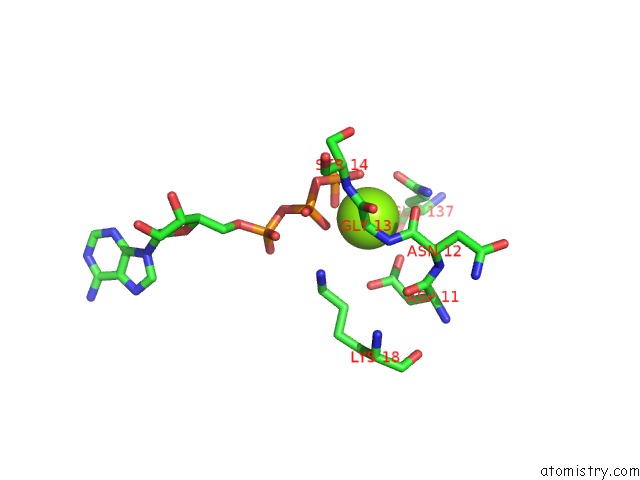
Magnesium binding site 2 out of 2 in 3u9d
Go back to
Magnesium binding site 2 out
of 2 in the Crystal Structure of A Chimera Containing the N-Terminal Domain (Residues 8-24) of Drosophila Ciboulot and the C-Terminal Domain (Residues 13-44) of Bovine Thymosin-BETA4, Bound to G-Actin-Atp
Mono view
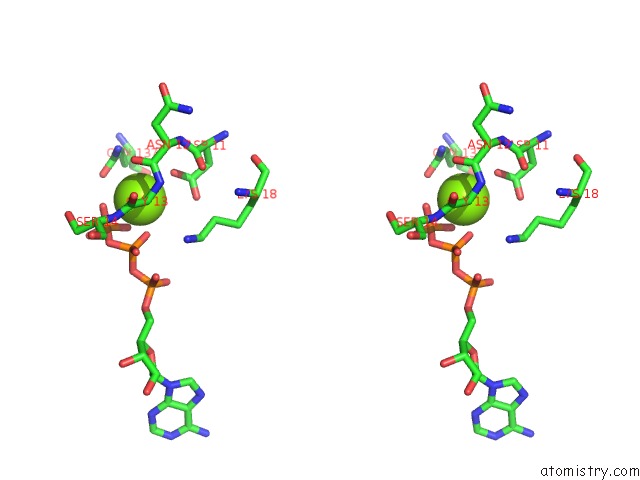
Stereo pair view
Mono view
Stereo pair view
A full contact list of Magnesium with other atoms in the Mg binding
site number 2 of Crystal Structure of A Chimera Containing the N-Terminal Domain (Residues 8-24) of Drosophila Ciboulot and the C-Terminal Domain (Residues 13-44) of Bovine Thymosin-BETA4, Bound to G-Actin-Atp within 5.0Å range:
|
Reference:
D.Didry,
F.X.Cantrelle,
C.Husson,
P.Roblin,
A.M.Moorthy,
J.Perez,
C.Le Clainche,
M.Hertzog,
E.Guittet,
M.F.Carlier,
C.Van Heijenoort,
L.Renault.
How A Single Residue in Individual Beta-Thymosin/WH2 Domains Controls Their Functions in Actin Assembly. Embo J. V. 31 1000 2012.
ISSN: ISSN 0261-4189
PubMed: 22193718
DOI: 10.1038/EMBOJ.2011.461
Page generated: Thu Aug 15 12:26:51 2024
ISSN: ISSN 0261-4189
PubMed: 22193718
DOI: 10.1038/EMBOJ.2011.461
Last articles
Fe in 2YXOFe in 2YRS
Fe in 2YXC
Fe in 2YNM
Fe in 2YVJ
Fe in 2YP1
Fe in 2YU2
Fe in 2YU1
Fe in 2YQB
Fe in 2YOO