Magnesium »
PDB 3umy-3v4i »
3uqd »
Magnesium in PDB 3uqd: Crystal Structure of the Phosphofructokinase-2 From Escherichia Coli in Complex with Substrates and Products
Enzymatic activity of Crystal Structure of the Phosphofructokinase-2 From Escherichia Coli in Complex with Substrates and Products
All present enzymatic activity of Crystal Structure of the Phosphofructokinase-2 From Escherichia Coli in Complex with Substrates and Products:
2.7.1.11;
2.7.1.11;
Protein crystallography data
The structure of Crystal Structure of the Phosphofructokinase-2 From Escherichia Coli in Complex with Substrates and Products, PDB code: 3uqd
was solved by
H.M.Pereira,
A.Caniuguir,
M.Baez,
R.Cabrera,
J.Babul,
with X-Ray Crystallography technique. A brief refinement statistics is given in the table below:
| Resolution Low / High (Å) | 19.79 / 2.14 |
| Space group | C 2 2 21 |
| Cell size a, b, c (Å), α, β, γ (°) | 69.885, 155.189, 224.989, 90.00, 90.00, 90.00 |
| R / Rfree (%) | 19.5 / 25.5 |
Magnesium Binding Sites:
The binding sites of Magnesium atom in the Crystal Structure of the Phosphofructokinase-2 From Escherichia Coli in Complex with Substrates and Products
(pdb code 3uqd). This binding sites where shown within
5.0 Angstroms radius around Magnesium atom.
In total 6 binding sites of Magnesium where determined in the Crystal Structure of the Phosphofructokinase-2 From Escherichia Coli in Complex with Substrates and Products, PDB code: 3uqd:
Jump to Magnesium binding site number: 1; 2; 3; 4; 5; 6;
In total 6 binding sites of Magnesium where determined in the Crystal Structure of the Phosphofructokinase-2 From Escherichia Coli in Complex with Substrates and Products, PDB code: 3uqd:
Jump to Magnesium binding site number: 1; 2; 3; 4; 5; 6;
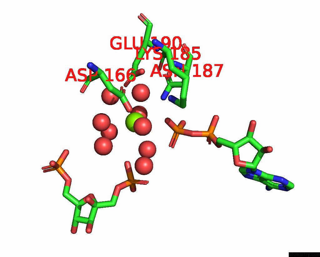
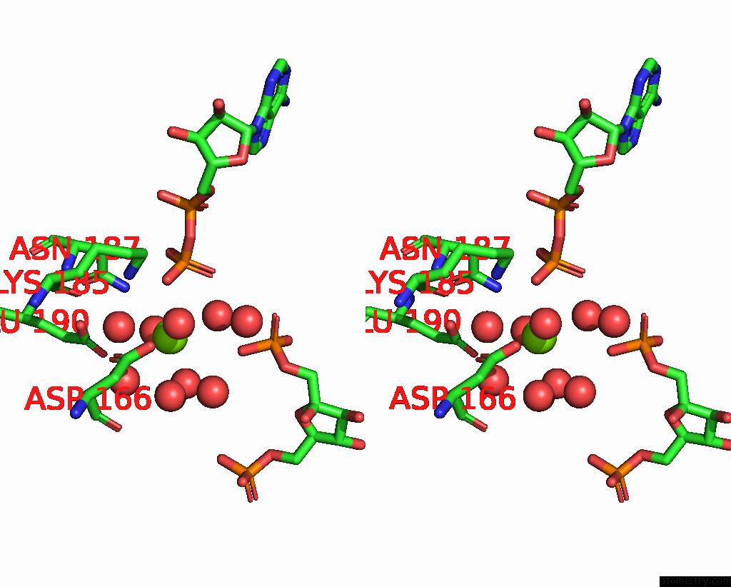
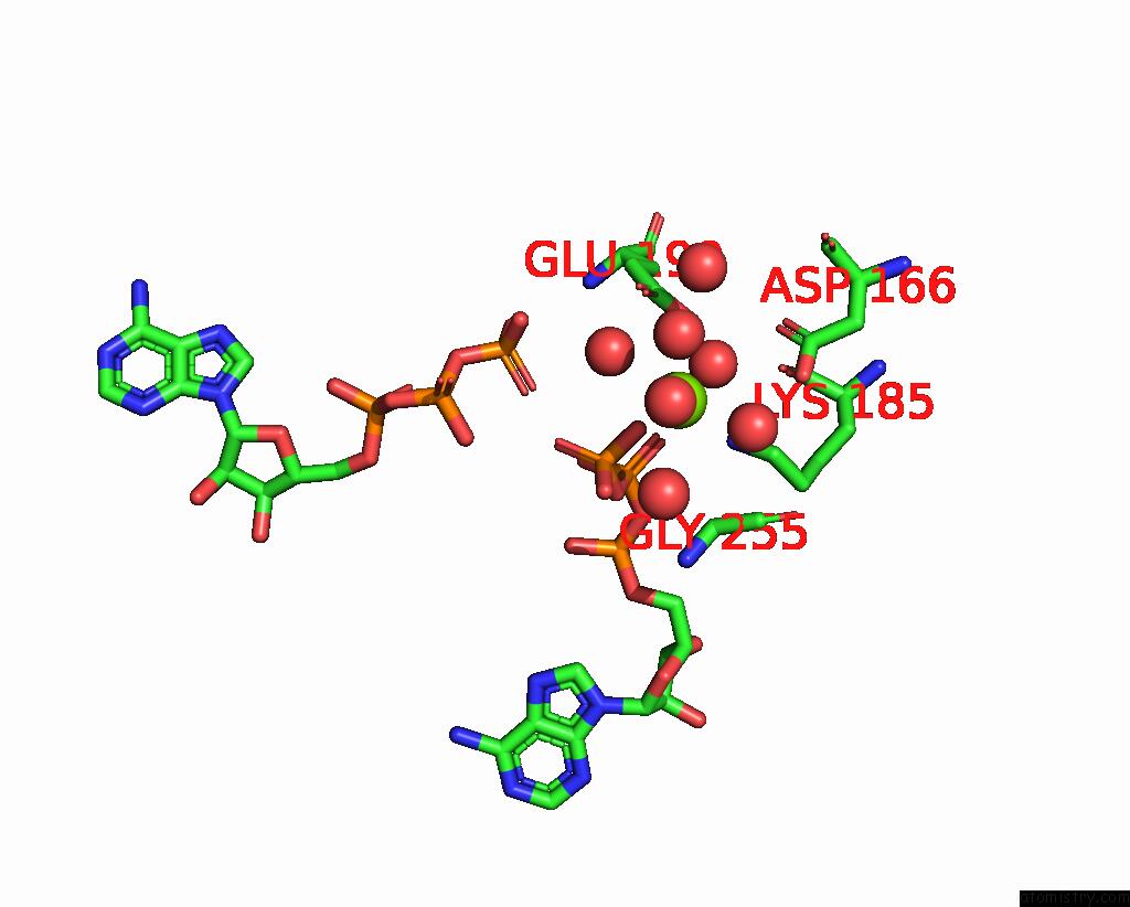
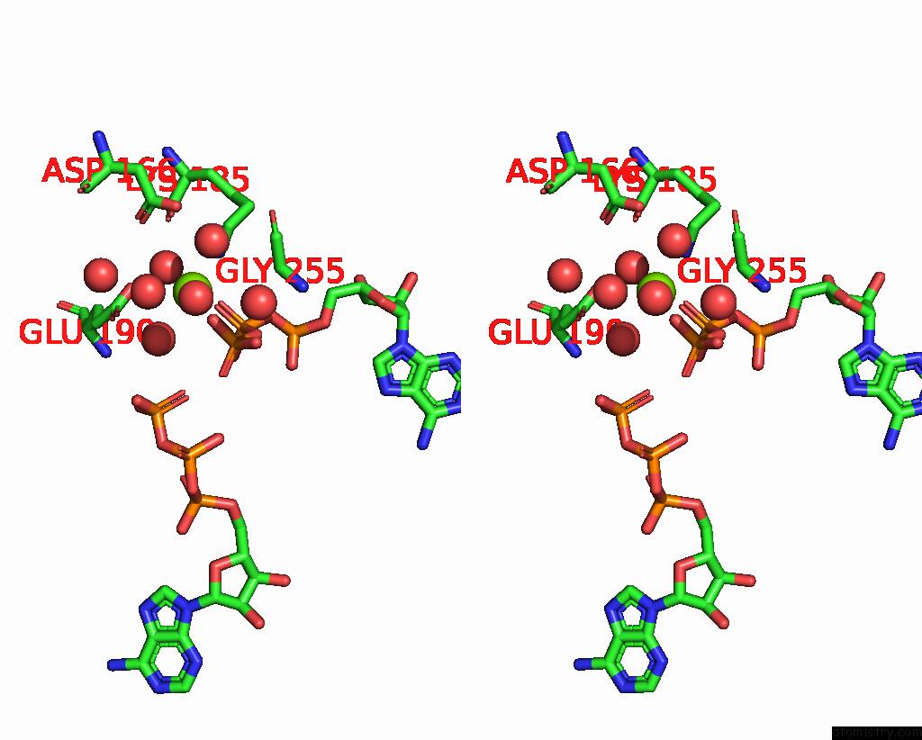
Magnesium binding site 1 out of 6 in 3uqd
Go back to
Magnesium binding site 1 out
of 6 in the Crystal Structure of the Phosphofructokinase-2 From Escherichia Coli in Complex with Substrates and Products
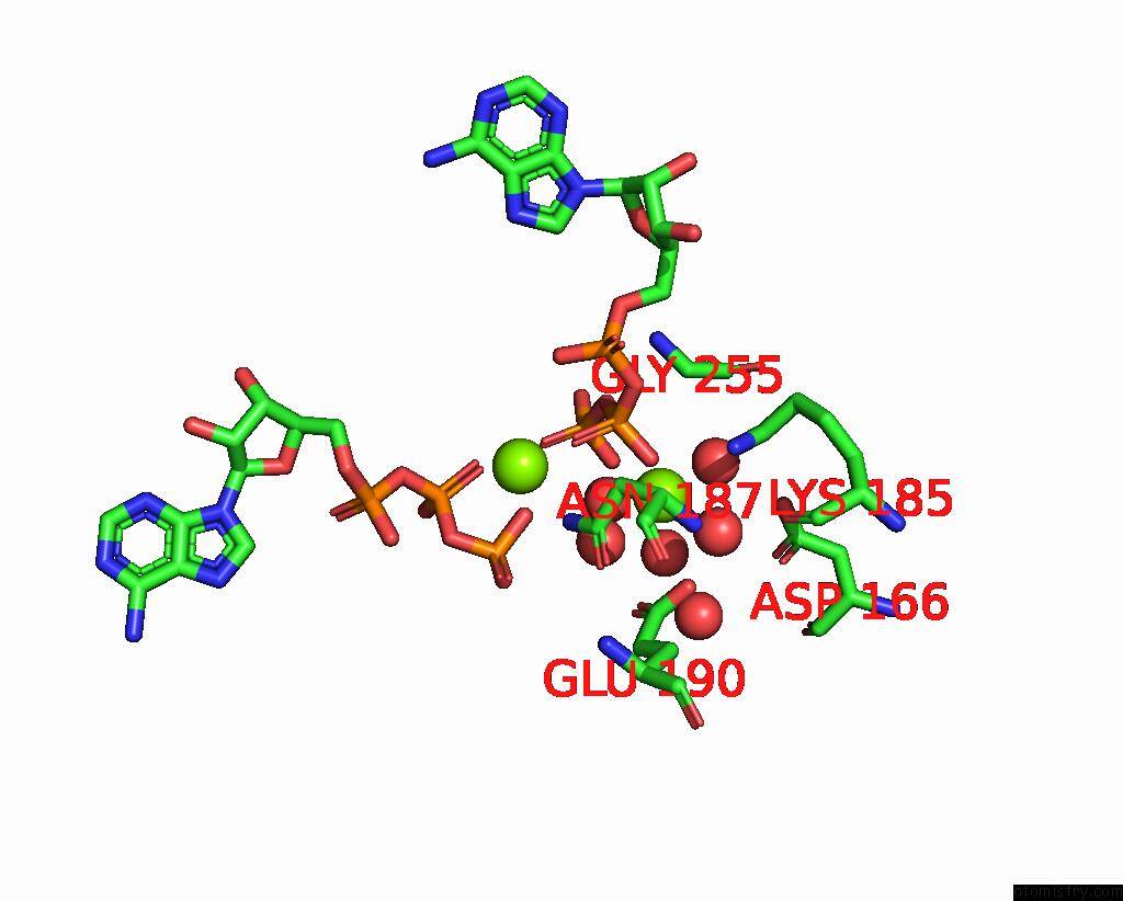
Mono view
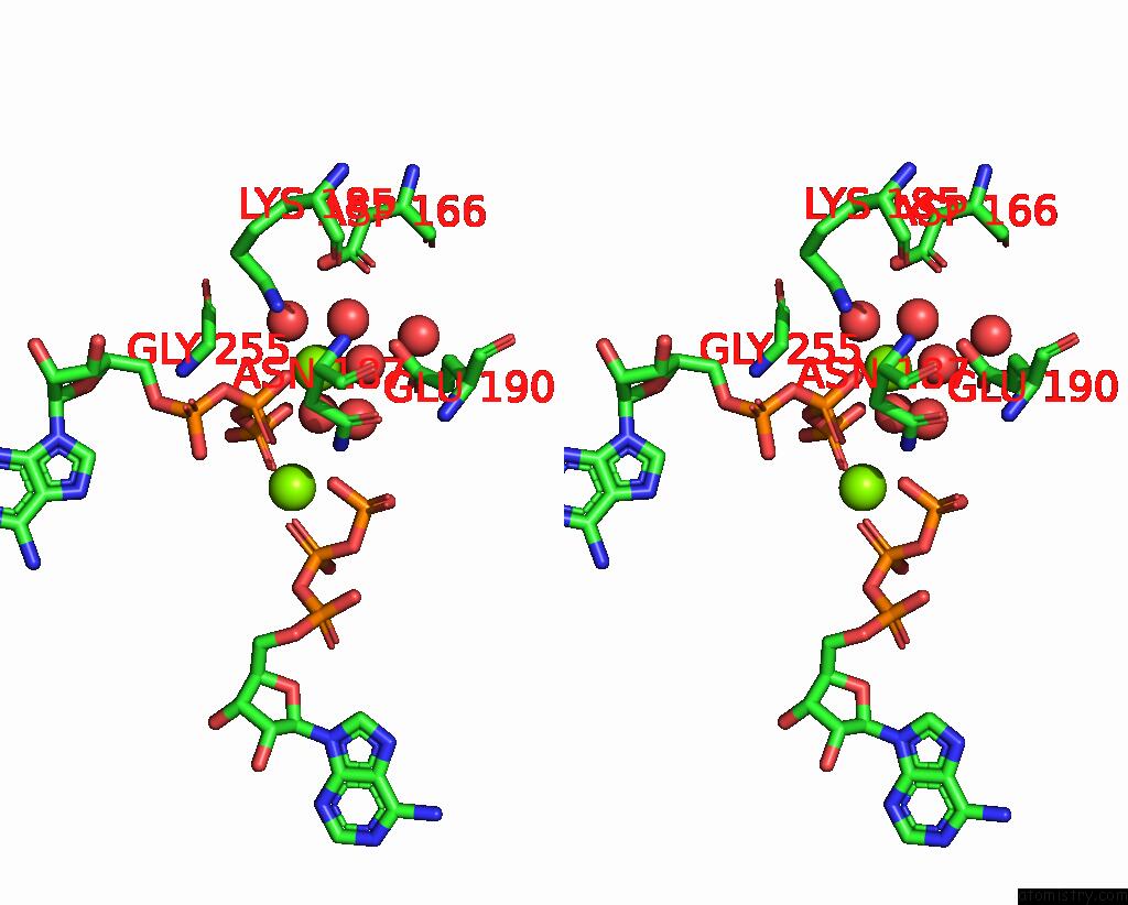
Stereo pair view
Mono view
Stereo pair view
A full contact list of Magnesium with other atoms in the Mg binding
site number 1 of Crystal Structure of the Phosphofructokinase-2 From Escherichia Coli in Complex with Substrates and Products within 5.0Å range:
|
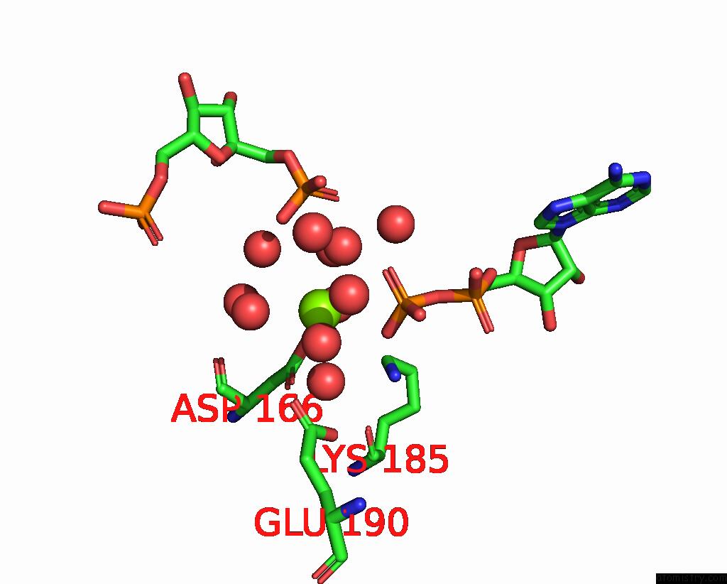
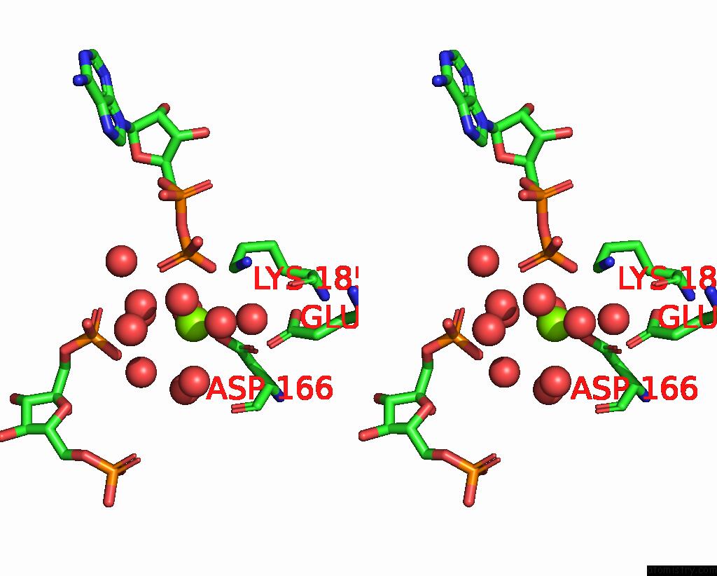
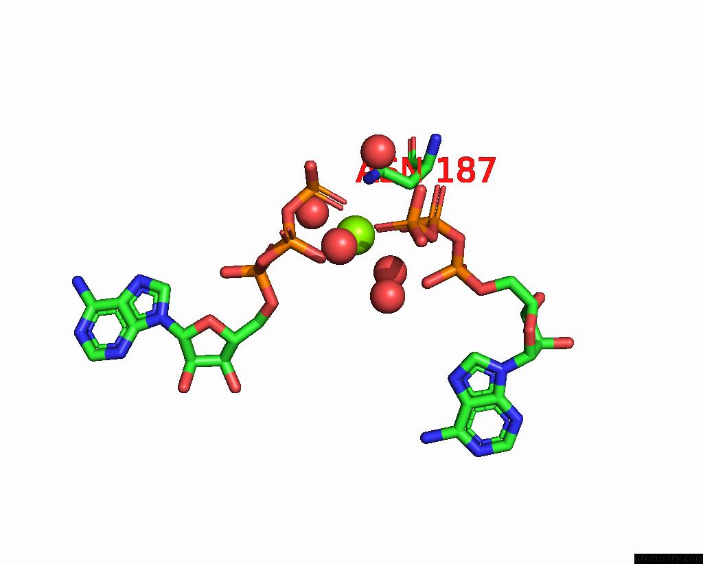
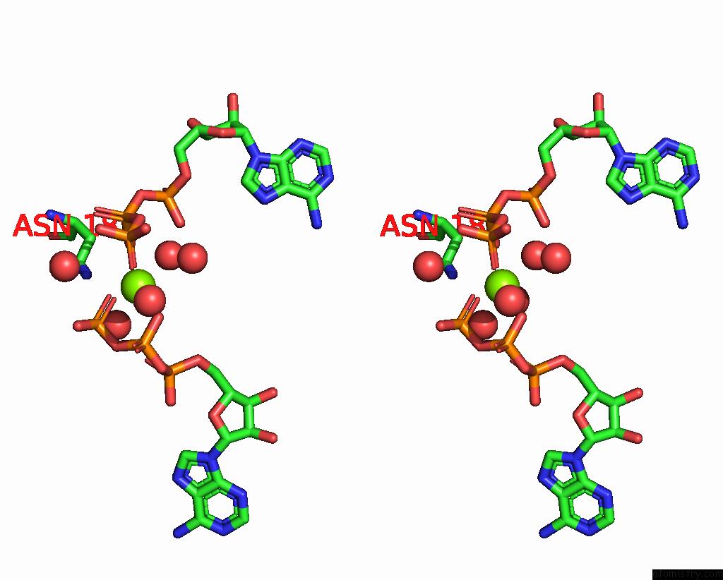
Magnesium binding site 2 out of 6 in 3uqd
Go back to
Magnesium binding site 2 out
of 6 in the Crystal Structure of the Phosphofructokinase-2 From Escherichia Coli in Complex with Substrates and Products
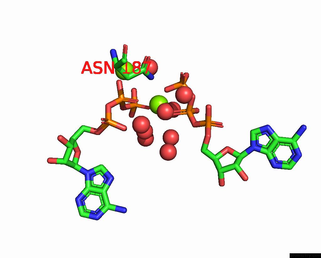
Mono view
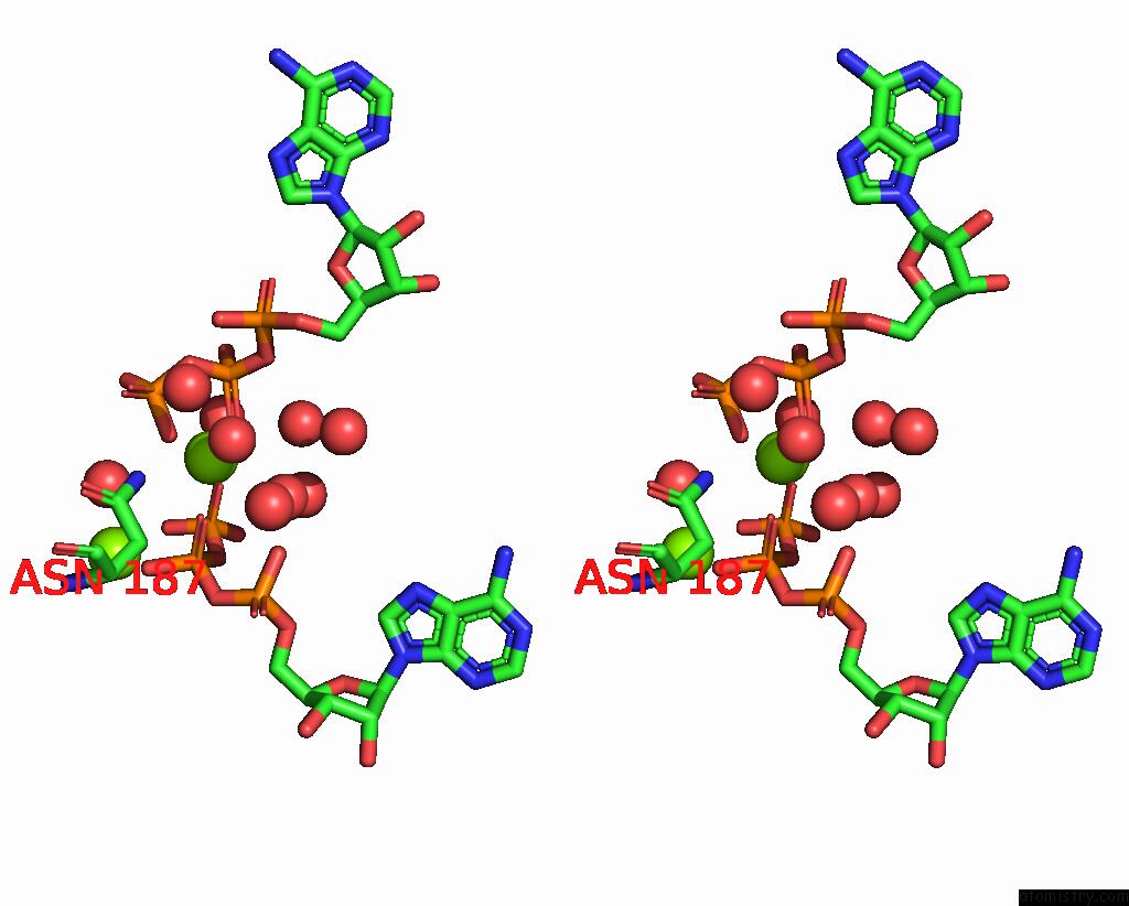
Stereo pair view
Mono view
Stereo pair view
A full contact list of Magnesium with other atoms in the Mg binding
site number 2 of Crystal Structure of the Phosphofructokinase-2 From Escherichia Coli in Complex with Substrates and Products within 5.0Å range:
|
Magnesium binding site 3 out of 6 in 3uqd
Go back to
Magnesium binding site 3 out
of 6 in the Crystal Structure of the Phosphofructokinase-2 From Escherichia Coli in Complex with Substrates and Products
Mono view
Stereo pair view
Mono view
Stereo pair view
A full contact list of Magnesium with other atoms in the Mg binding
site number 3 of Crystal Structure of the Phosphofructokinase-2 From Escherichia Coli in Complex with Substrates and Products within 5.0Å range:
|
Magnesium binding site 4 out of 6 in 3uqd
Go back to
Magnesium binding site 4 out
of 6 in the Crystal Structure of the Phosphofructokinase-2 From Escherichia Coli in Complex with Substrates and Products
Mono view
Stereo pair view
Mono view
Stereo pair view
A full contact list of Magnesium with other atoms in the Mg binding
site number 4 of Crystal Structure of the Phosphofructokinase-2 From Escherichia Coli in Complex with Substrates and Products within 5.0Å range:
|
Magnesium binding site 5 out of 6 in 3uqd
Go back to
Magnesium binding site 5 out
of 6 in the Crystal Structure of the Phosphofructokinase-2 From Escherichia Coli in Complex with Substrates and Products
Mono view
Stereo pair view
Mono view
Stereo pair view
A full contact list of Magnesium with other atoms in the Mg binding
site number 5 of Crystal Structure of the Phosphofructokinase-2 From Escherichia Coli in Complex with Substrates and Products within 5.0Å range:
|
Magnesium binding site 6 out of 6 in 3uqd
Go back to
Magnesium binding site 6 out
of 6 in the Crystal Structure of the Phosphofructokinase-2 From Escherichia Coli in Complex with Substrates and Products
Mono view
Stereo pair view
Mono view
Stereo pair view
A full contact list of Magnesium with other atoms in the Mg binding
site number 6 of Crystal Structure of the Phosphofructokinase-2 From Escherichia Coli in Complex with Substrates and Products within 5.0Å range:
|
Reference:
J.Murillo-Lopez,
K.Zinovjev,
H.Pereira,
A.Caniuguir,
R.Garratt,
J.Babul,
R.Recabarren,
J.Alzate-Morales,
J.Caballero,
I.Tunon,
R.Cabrera.
Studying the Phosphoryl Transfer Mechanism of Thee. Coliphosphofructokinase-2: From X-Ray Structure to Quantum Mechanics/Molecular Mechanics Simulations. Chem Sci V. 10 2882 2019.
ISSN: ISSN 2041-6520
PubMed: 30996866
DOI: 10.1039/C9SC00094A
Page generated: Thu Aug 15 12:37:38 2024
ISSN: ISSN 2041-6520
PubMed: 30996866
DOI: 10.1039/C9SC00094A
Last articles
Zn in 9MJ5Zn in 9HNW
Zn in 9G0L
Zn in 9FNE
Zn in 9DZN
Zn in 9E0I
Zn in 9D32
Zn in 9DAK
Zn in 8ZXC
Zn in 8ZUF