Magnesium »
PDB 3ump-3v4f »
3uul »
Magnesium in PDB 3uul: Crystal Structure of First N-Terminal Utrophin Spectrin Repeat
Protein crystallography data
The structure of Crystal Structure of First N-Terminal Utrophin Spectrin Repeat, PDB code: 3uul
was solved by
M.Muthu,
K.A.Richardson,
A.J.Sutherland-Smith,
with X-Ray Crystallography technique. A brief refinement statistics is given in the table below:
| Resolution Low / High (Å) | 27.63 / 1.95 |
| Space group | P 21 21 21 |
| Cell size a, b, c (Å), α, β, γ (°) | 43.000, 58.660, 91.450, 90.00, 90.00, 90.00 |
| R / Rfree (%) | 19.8 / 23.5 |
Magnesium Binding Sites:
The binding sites of Magnesium atom in the Crystal Structure of First N-Terminal Utrophin Spectrin Repeat
(pdb code 3uul). This binding sites where shown within
5.0 Angstroms radius around Magnesium atom.
In total only one binding site of Magnesium was determined in the Crystal Structure of First N-Terminal Utrophin Spectrin Repeat, PDB code: 3uul:
In total only one binding site of Magnesium was determined in the Crystal Structure of First N-Terminal Utrophin Spectrin Repeat, PDB code: 3uul:
Magnesium binding site 1 out of 1 in 3uul
Go back to
Magnesium binding site 1 out
of 1 in the Crystal Structure of First N-Terminal Utrophin Spectrin Repeat
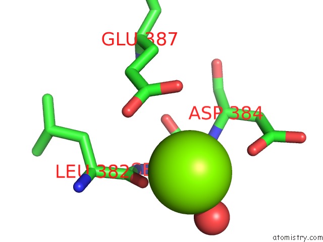
Mono view
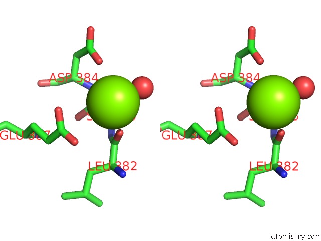
Stereo pair view
Mono view
Stereo pair view
A full contact list of Magnesium with other atoms in the Mg binding
site number 1 of Crystal Structure of First N-Terminal Utrophin Spectrin Repeat within 5.0Å range:
|
Reference:
M.Muthu,
K.A.Richardson,
A.J.Sutherland-Smith.
The Crystal Structures of Dystrophin and Utrophin Spectrin Repeats: Implications For Domain Boundaries Plos One V. 7 40066 2012.
ISSN: ESSN 1932-6203
PubMed: 22911693
DOI: 10.1371/JOURNAL.PONE.0040066
Page generated: Thu Aug 15 12:39:08 2024
ISSN: ESSN 1932-6203
PubMed: 22911693
DOI: 10.1371/JOURNAL.PONE.0040066
Last articles
Zn in 9J0NZn in 9J0O
Zn in 9J0P
Zn in 9FJX
Zn in 9EKB
Zn in 9C0F
Zn in 9CAH
Zn in 9CH0
Zn in 9CH3
Zn in 9CH1