Magnesium »
PDB 3ump-3v4f »
3uyl »
Magnesium in PDB 3uyl: Spinosyn Rhamnosyltransferase Spng Complexed with Thymidine Diphosphate
Protein crystallography data
The structure of Spinosyn Rhamnosyltransferase Spng Complexed with Thymidine Diphosphate, PDB code: 3uyl
was solved by
E.A.Isiorho,
H.-W.Liu,
A.T.Keatinge-Clay,
with X-Ray Crystallography technique. A brief refinement statistics is given in the table below:
| Resolution Low / High (Å) | 24.62 / 1.85 |
| Space group | P 1 |
| Cell size a, b, c (Å), α, β, γ (°) | 54.152, 57.629, 68.203, 81.63, 73.79, 85.74 |
| R / Rfree (%) | 23.2 / 29.1 |
Magnesium Binding Sites:
The binding sites of Magnesium atom in the Spinosyn Rhamnosyltransferase Spng Complexed with Thymidine Diphosphate
(pdb code 3uyl). This binding sites where shown within
5.0 Angstroms radius around Magnesium atom.
In total 2 binding sites of Magnesium where determined in the Spinosyn Rhamnosyltransferase Spng Complexed with Thymidine Diphosphate, PDB code: 3uyl:
Jump to Magnesium binding site number: 1; 2;
In total 2 binding sites of Magnesium where determined in the Spinosyn Rhamnosyltransferase Spng Complexed with Thymidine Diphosphate, PDB code: 3uyl:
Jump to Magnesium binding site number: 1; 2;
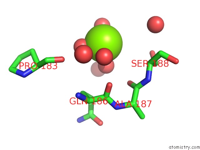
Magnesium binding site 1 out of 2 in 3uyl
Go back to
Magnesium binding site 1 out
of 2 in the Spinosyn Rhamnosyltransferase Spng Complexed with Thymidine Diphosphate
Mono view
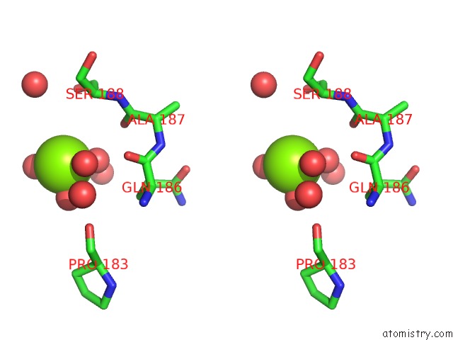
Stereo pair view
Mono view
Stereo pair view
A full contact list of Magnesium with other atoms in the Mg binding
site number 1 of Spinosyn Rhamnosyltransferase Spng Complexed with Thymidine Diphosphate within 5.0Å range:
|
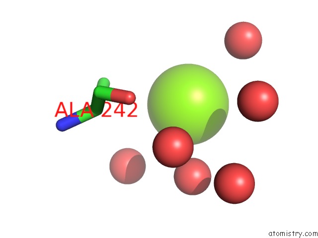
Magnesium binding site 2 out of 2 in 3uyl
Go back to
Magnesium binding site 2 out
of 2 in the Spinosyn Rhamnosyltransferase Spng Complexed with Thymidine Diphosphate
Mono view
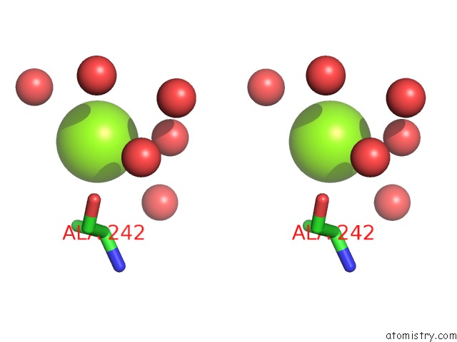
Stereo pair view
Mono view
Stereo pair view
A full contact list of Magnesium with other atoms in the Mg binding
site number 2 of Spinosyn Rhamnosyltransferase Spng Complexed with Thymidine Diphosphate within 5.0Å range:
|
Reference:
E.A.Isiorho,
H.W.Liu,
A.T.Keatinge-Clay.
Structural Studies of the Spinosyn Rhamnosyltransferase, Spng. Biochemistry V. 51 1213 2012.
ISSN: ISSN 0006-2960
PubMed: 22283226
DOI: 10.1021/BI201860Q
Page generated: Thu Aug 15 12:41:18 2024
ISSN: ISSN 0006-2960
PubMed: 22283226
DOI: 10.1021/BI201860Q
Last articles
Zn in 9J0NZn in 9J0O
Zn in 9J0P
Zn in 9FJX
Zn in 9EKB
Zn in 9C0F
Zn in 9CAH
Zn in 9CH0
Zn in 9CH3
Zn in 9CH1