Magnesium »
PDB 3ump-3v4f »
3uzt »
Magnesium in PDB 3uzt: Structure of the C13.18 Rna Aptamer in Complex with G Protein-Coupled Receptor Kinase 2
Enzymatic activity of Structure of the C13.18 Rna Aptamer in Complex with G Protein-Coupled Receptor Kinase 2
All present enzymatic activity of Structure of the C13.18 Rna Aptamer in Complex with G Protein-Coupled Receptor Kinase 2:
2.7.11.15;
2.7.11.15;
Protein crystallography data
The structure of Structure of the C13.18 Rna Aptamer in Complex with G Protein-Coupled Receptor Kinase 2, PDB code: 3uzt
was solved by
J.J.G.Tesmer,
V.M.Tesmer,
with X-Ray Crystallography technique. A brief refinement statistics is given in the table below:
| Resolution Low / High (Å) | 24.88 / 3.51 |
| Space group | P 21 21 2 |
| Cell size a, b, c (Å), α, β, γ (°) | 113.346, 139.719, 60.949, 90.00, 90.00, 90.00 |
| R / Rfree (%) | 21.6 / 31.7 |
Magnesium Binding Sites:
The binding sites of Magnesium atom in the Structure of the C13.18 Rna Aptamer in Complex with G Protein-Coupled Receptor Kinase 2
(pdb code 3uzt). This binding sites where shown within
5.0 Angstroms radius around Magnesium atom.
In total only one binding site of Magnesium was determined in the Structure of the C13.18 Rna Aptamer in Complex with G Protein-Coupled Receptor Kinase 2, PDB code: 3uzt:
In total only one binding site of Magnesium was determined in the Structure of the C13.18 Rna Aptamer in Complex with G Protein-Coupled Receptor Kinase 2, PDB code: 3uzt:
Magnesium binding site 1 out of 1 in 3uzt
Go back to
Magnesium binding site 1 out
of 1 in the Structure of the C13.18 Rna Aptamer in Complex with G Protein-Coupled Receptor Kinase 2
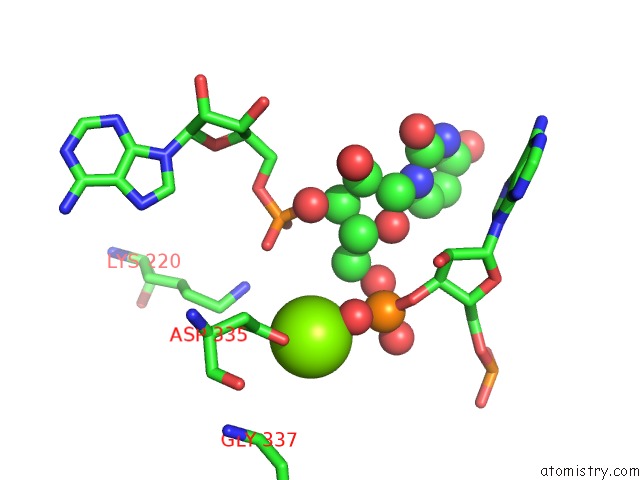
Mono view
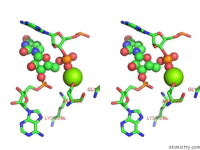
Stereo pair view
Mono view
Stereo pair view
A full contact list of Magnesium with other atoms in the Mg binding
site number 1 of Structure of the C13.18 Rna Aptamer in Complex with G Protein-Coupled Receptor Kinase 2 within 5.0Å range:
|
Reference:
V.M.Tesmer,
S.Lennarz,
G.Mayer,
J.J.Tesmer.
Molecular Mechanism For Inhibition of G Protein-Coupled Receptor Kinase 2 By A Selective Rna Aptamer. Structure V. 20 1300 2012.
ISSN: ISSN 0969-2126
PubMed: 22727813
DOI: 10.1016/J.STR.2012.05.002
Page generated: Thu Aug 15 12:42:04 2024
ISSN: ISSN 0969-2126
PubMed: 22727813
DOI: 10.1016/J.STR.2012.05.002
Last articles
Zn in 9J0NZn in 9J0O
Zn in 9J0P
Zn in 9FJX
Zn in 9EKB
Zn in 9C0F
Zn in 9CAH
Zn in 9CH0
Zn in 9CH3
Zn in 9CH1