Magnesium »
PDB 3v4k-3vdb »
3v7e »
Magnesium in PDB 3v7e: Crystal Structure of Ybxf Bound to the Sam-I Riboswitch Aptamer
Protein crystallography data
The structure of Crystal Structure of Ybxf Bound to the Sam-I Riboswitch Aptamer, PDB code: 3v7e
was solved by
N.J.Baird,
J.Zhang,
T.Hamma,
A.R.Ferre-D'amare,
with X-Ray Crystallography technique. A brief refinement statistics is given in the table below:
| Resolution Low / High (Å) | 29.54 / 2.80 |
| Space group | C 1 2 1 |
| Cell size a, b, c (Å), α, β, γ (°) | 191.762, 54.305, 106.366, 90.00, 116.56, 90.00 |
| R / Rfree (%) | 21.8 / 27.4 |
Other elements in 3v7e:
The structure of Crystal Structure of Ybxf Bound to the Sam-I Riboswitch Aptamer also contains other interesting chemical elements:
| Cobalt | (Co) | 18 atoms |
Magnesium Binding Sites:
Pages:
>>> Page 1 <<< Page 2, Binding sites: 11 - 20; Page 3, Binding sites: 21 - 22;Binding sites:
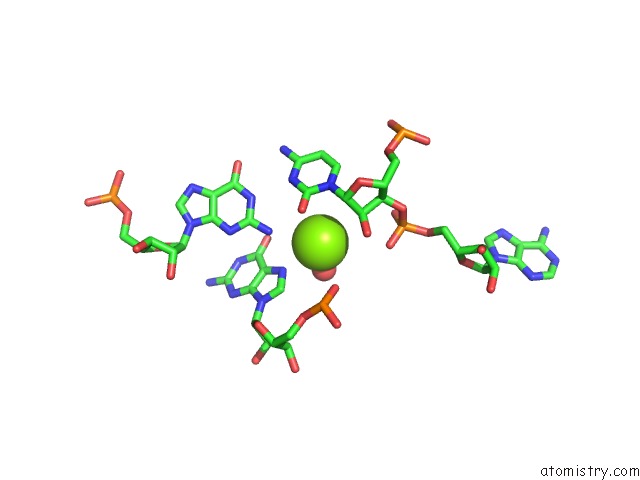
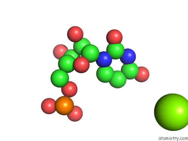
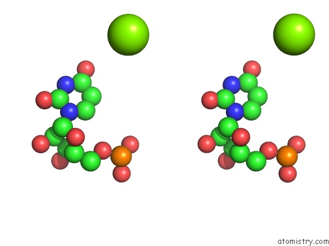
The binding sites of Magnesium atom in the Crystal Structure of Ybxf Bound to the Sam-I Riboswitch Aptamer (pdb code 3v7e). This binding sites where shown within 5.0 Angstroms radius around Magnesium atom.In total 22 binding sites of Magnesium where determined in the Crystal Structure of Ybxf Bound to the Sam-I Riboswitch Aptamer, PDB code: 3v7e:
Jump to Magnesium binding site number: 1; 2; 3; 4; 5; 6; 7; 8; 9; 10;
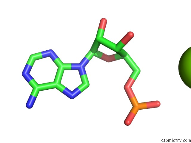
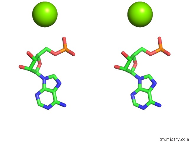
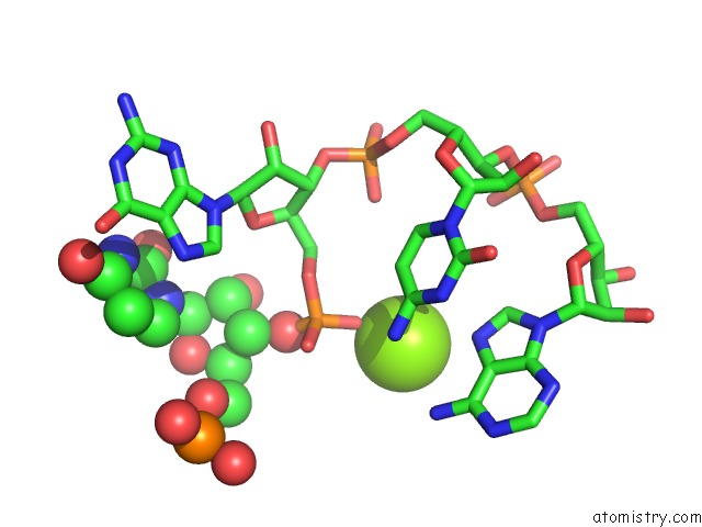
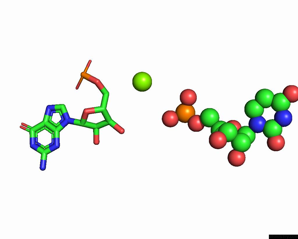
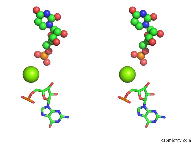
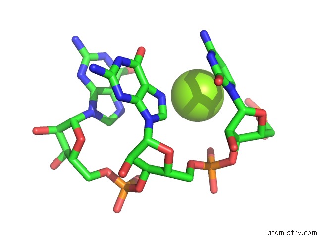
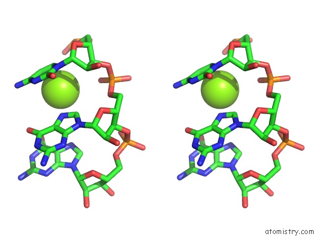
Magnesium binding site 1 out of 22 in 3v7e
Go back to
Magnesium binding site 1 out
of 22 in the Crystal Structure of Ybxf Bound to the Sam-I Riboswitch Aptamer
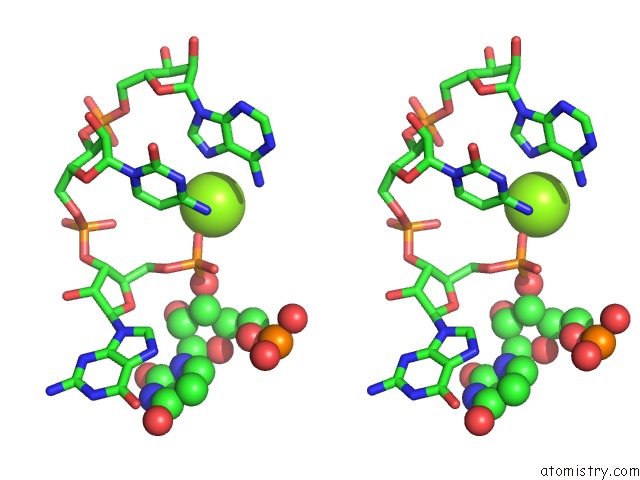
Mono view
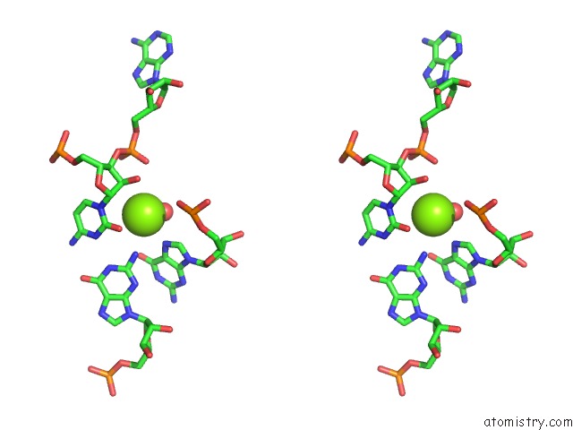
Stereo pair view
Mono view
Stereo pair view
A full contact list of Magnesium with other atoms in the Mg binding
site number 1 of Crystal Structure of Ybxf Bound to the Sam-I Riboswitch Aptamer within 5.0Å range:
|
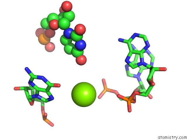
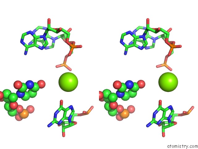
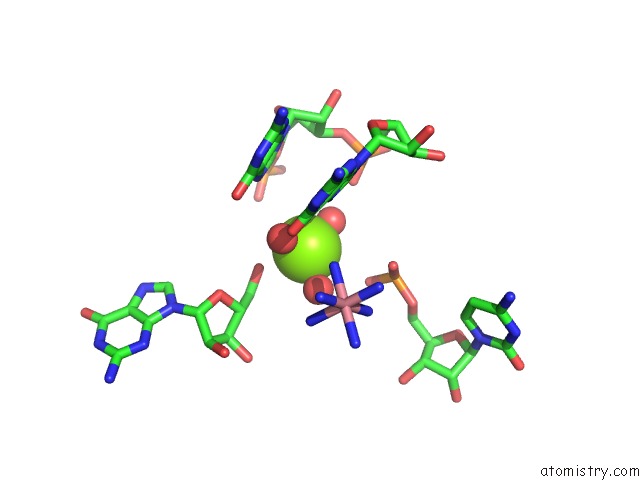
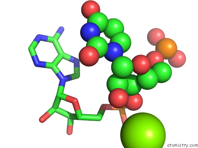
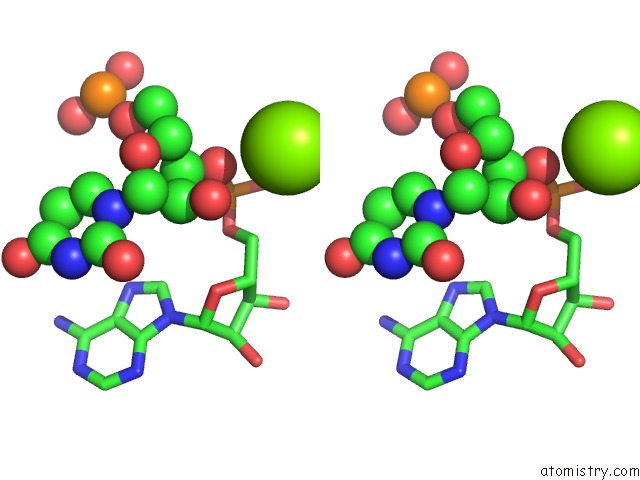
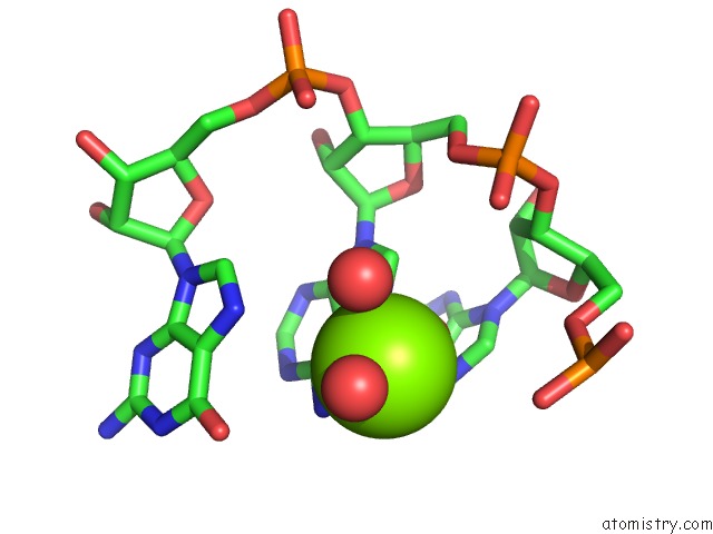
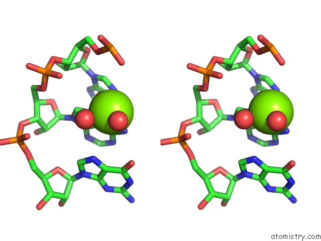
Magnesium binding site 2 out of 22 in 3v7e
Go back to
Magnesium binding site 2 out
of 22 in the Crystal Structure of Ybxf Bound to the Sam-I Riboswitch Aptamer
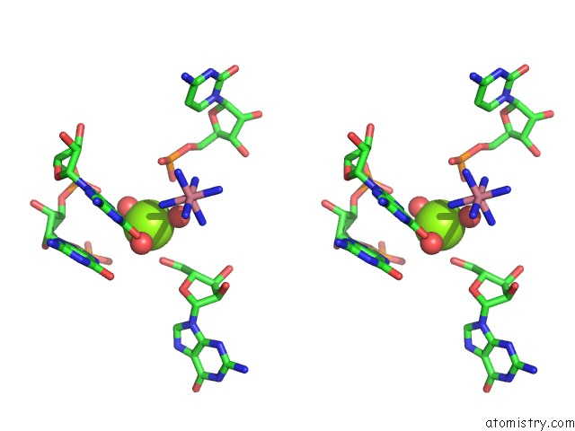
Mono view
Stereo pair view
Mono view
Stereo pair view
A full contact list of Magnesium with other atoms in the Mg binding
site number 2 of Crystal Structure of Ybxf Bound to the Sam-I Riboswitch Aptamer within 5.0Å range:
|
Magnesium binding site 3 out of 22 in 3v7e
Go back to
Magnesium binding site 3 out
of 22 in the Crystal Structure of Ybxf Bound to the Sam-I Riboswitch Aptamer
Mono view
Stereo pair view
Mono view
Stereo pair view
A full contact list of Magnesium with other atoms in the Mg binding
site number 3 of Crystal Structure of Ybxf Bound to the Sam-I Riboswitch Aptamer within 5.0Å range:
|
Magnesium binding site 4 out of 22 in 3v7e
Go back to
Magnesium binding site 4 out
of 22 in the Crystal Structure of Ybxf Bound to the Sam-I Riboswitch Aptamer
Mono view
Stereo pair view
Mono view
Stereo pair view
A full contact list of Magnesium with other atoms in the Mg binding
site number 4 of Crystal Structure of Ybxf Bound to the Sam-I Riboswitch Aptamer within 5.0Å range:
|
Magnesium binding site 5 out of 22 in 3v7e
Go back to
Magnesium binding site 5 out
of 22 in the Crystal Structure of Ybxf Bound to the Sam-I Riboswitch Aptamer
Mono view
Stereo pair view
Mono view
Stereo pair view
A full contact list of Magnesium with other atoms in the Mg binding
site number 5 of Crystal Structure of Ybxf Bound to the Sam-I Riboswitch Aptamer within 5.0Å range:
|
Magnesium binding site 6 out of 22 in 3v7e
Go back to
Magnesium binding site 6 out
of 22 in the Crystal Structure of Ybxf Bound to the Sam-I Riboswitch Aptamer
Mono view
Stereo pair view
Mono view
Stereo pair view
A full contact list of Magnesium with other atoms in the Mg binding
site number 6 of Crystal Structure of Ybxf Bound to the Sam-I Riboswitch Aptamer within 5.0Å range:
|
Magnesium binding site 7 out of 22 in 3v7e
Go back to
Magnesium binding site 7 out
of 22 in the Crystal Structure of Ybxf Bound to the Sam-I Riboswitch Aptamer
Mono view
Stereo pair view
Mono view
Stereo pair view
A full contact list of Magnesium with other atoms in the Mg binding
site number 7 of Crystal Structure of Ybxf Bound to the Sam-I Riboswitch Aptamer within 5.0Å range:
|
Magnesium binding site 8 out of 22 in 3v7e
Go back to
Magnesium binding site 8 out
of 22 in the Crystal Structure of Ybxf Bound to the Sam-I Riboswitch Aptamer
Mono view
Stereo pair view
Mono view
Stereo pair view
A full contact list of Magnesium with other atoms in the Mg binding
site number 8 of Crystal Structure of Ybxf Bound to the Sam-I Riboswitch Aptamer within 5.0Å range:
|
Magnesium binding site 9 out of 22 in 3v7e
Go back to
Magnesium binding site 9 out
of 22 in the Crystal Structure of Ybxf Bound to the Sam-I Riboswitch Aptamer
Mono view
Stereo pair view
Mono view
Stereo pair view
A full contact list of Magnesium with other atoms in the Mg binding
site number 9 of Crystal Structure of Ybxf Bound to the Sam-I Riboswitch Aptamer within 5.0Å range:
|
Magnesium binding site 10 out of 22 in 3v7e
Go back to
Magnesium binding site 10 out
of 22 in the Crystal Structure of Ybxf Bound to the Sam-I Riboswitch Aptamer
Mono view
Stereo pair view
Mono view
Stereo pair view
A full contact list of Magnesium with other atoms in the Mg binding
site number 10 of Crystal Structure of Ybxf Bound to the Sam-I Riboswitch Aptamer within 5.0Å range:
|
Reference:
N.J.Baird,
J.Zhang,
T.Hamma,
A.R.Ferre-D'amare.
Ybxf and Ylxq Are Bacterial Homologs of L7AE and Bind K-Turns But Not K-Loops. Rna V. 18 759 2012.
ISSN: ISSN 1355-8382
PubMed: 22355167
DOI: 10.1261/RNA.031518.111
Page generated: Thu Aug 15 12:45:53 2024
ISSN: ISSN 1355-8382
PubMed: 22355167
DOI: 10.1261/RNA.031518.111
Last articles
Fe in 2YXOFe in 2YRS
Fe in 2YXC
Fe in 2YNM
Fe in 2YVJ
Fe in 2YP1
Fe in 2YU2
Fe in 2YU1
Fe in 2YQB
Fe in 2YOO