Magnesium »
PDB 3v4k-3vdb »
3vc2 »
Magnesium in PDB 3vc2: Crystal Structure of Geranyl Diphosphate C-Methyltransferase From Streptomyces Coelicolor A3(2) in Complex with MG2+, Geranyl Diphosphate, and S-Adenosyl-L-Homocysteine
Protein crystallography data
The structure of Crystal Structure of Geranyl Diphosphate C-Methyltransferase From Streptomyces Coelicolor A3(2) in Complex with MG2+, Geranyl Diphosphate, and S-Adenosyl-L-Homocysteine, PDB code: 3vc2
was solved by
M.Koksal,
D.W.Christianson,
with X-Ray Crystallography technique. A brief refinement statistics is given in the table below:
| Resolution Low / High (Å) | 48.89 / 2.05 |
| Space group | P 1 21 1 |
| Cell size a, b, c (Å), α, β, γ (°) | 98.128, 103.252, 204.132, 90.00, 99.05, 90.00 |
| R / Rfree (%) | 18.1 / 22.2 |
Magnesium Binding Sites:
The binding sites of Magnesium atom in the Crystal Structure of Geranyl Diphosphate C-Methyltransferase From Streptomyces Coelicolor A3(2) in Complex with MG2+, Geranyl Diphosphate, and S-Adenosyl-L-Homocysteine
(pdb code 3vc2). This binding sites where shown within
5.0 Angstroms radius around Magnesium atom.
In total 4 binding sites of Magnesium where determined in the Crystal Structure of Geranyl Diphosphate C-Methyltransferase From Streptomyces Coelicolor A3(2) in Complex with MG2+, Geranyl Diphosphate, and S-Adenosyl-L-Homocysteine, PDB code: 3vc2:
Jump to Magnesium binding site number: 1; 2; 3; 4;
In total 4 binding sites of Magnesium where determined in the Crystal Structure of Geranyl Diphosphate C-Methyltransferase From Streptomyces Coelicolor A3(2) in Complex with MG2+, Geranyl Diphosphate, and S-Adenosyl-L-Homocysteine, PDB code: 3vc2:
Jump to Magnesium binding site number: 1; 2; 3; 4;
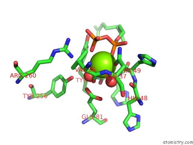
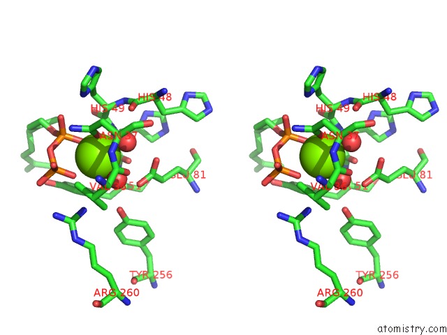
Magnesium binding site 1 out of 4 in 3vc2
Go back to
Magnesium binding site 1 out
of 4 in the Crystal Structure of Geranyl Diphosphate C-Methyltransferase From Streptomyces Coelicolor A3(2) in Complex with MG2+, Geranyl Diphosphate, and S-Adenosyl-L-Homocysteine
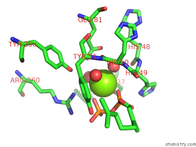
Mono view
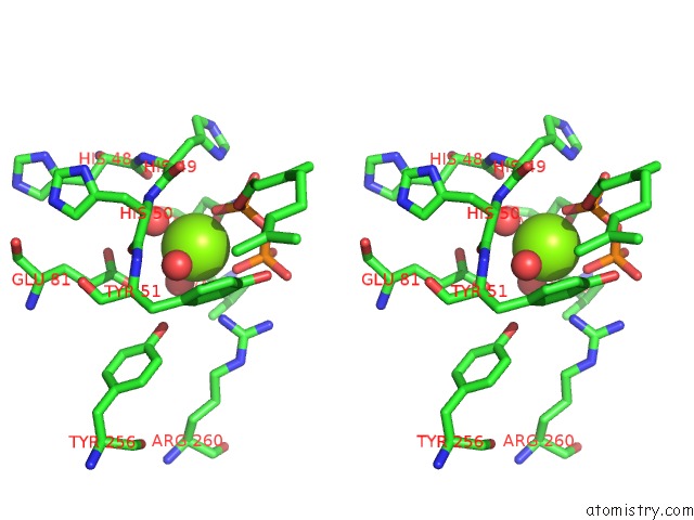
Stereo pair view
Mono view
Stereo pair view
A full contact list of Magnesium with other atoms in the Mg binding
site number 1 of Crystal Structure of Geranyl Diphosphate C-Methyltransferase From Streptomyces Coelicolor A3(2) in Complex with MG2+, Geranyl Diphosphate, and S-Adenosyl-L-Homocysteine within 5.0Å range:
|
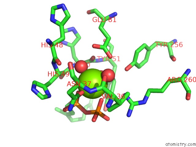
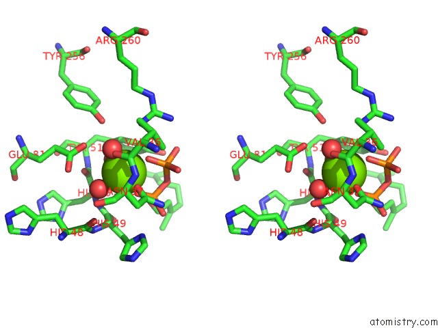
Magnesium binding site 2 out of 4 in 3vc2
Go back to
Magnesium binding site 2 out
of 4 in the Crystal Structure of Geranyl Diphosphate C-Methyltransferase From Streptomyces Coelicolor A3(2) in Complex with MG2+, Geranyl Diphosphate, and S-Adenosyl-L-Homocysteine
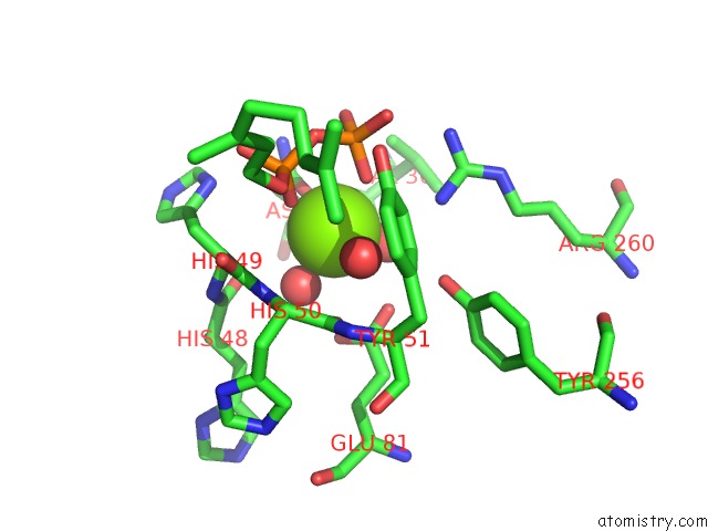
Mono view
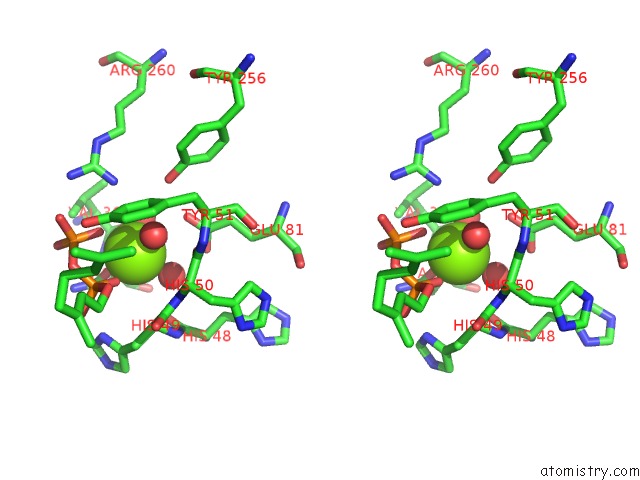
Stereo pair view
Mono view
Stereo pair view
A full contact list of Magnesium with other atoms in the Mg binding
site number 2 of Crystal Structure of Geranyl Diphosphate C-Methyltransferase From Streptomyces Coelicolor A3(2) in Complex with MG2+, Geranyl Diphosphate, and S-Adenosyl-L-Homocysteine within 5.0Å range:
|
Magnesium binding site 3 out of 4 in 3vc2
Go back to
Magnesium binding site 3 out
of 4 in the Crystal Structure of Geranyl Diphosphate C-Methyltransferase From Streptomyces Coelicolor A3(2) in Complex with MG2+, Geranyl Diphosphate, and S-Adenosyl-L-Homocysteine
Mono view
Stereo pair view
Mono view
Stereo pair view
A full contact list of Magnesium with other atoms in the Mg binding
site number 3 of Crystal Structure of Geranyl Diphosphate C-Methyltransferase From Streptomyces Coelicolor A3(2) in Complex with MG2+, Geranyl Diphosphate, and S-Adenosyl-L-Homocysteine within 5.0Å range:
|
Magnesium binding site 4 out of 4 in 3vc2
Go back to
Magnesium binding site 4 out
of 4 in the Crystal Structure of Geranyl Diphosphate C-Methyltransferase From Streptomyces Coelicolor A3(2) in Complex with MG2+, Geranyl Diphosphate, and S-Adenosyl-L-Homocysteine
Mono view
Stereo pair view
Mono view
Stereo pair view
A full contact list of Magnesium with other atoms in the Mg binding
site number 4 of Crystal Structure of Geranyl Diphosphate C-Methyltransferase From Streptomyces Coelicolor A3(2) in Complex with MG2+, Geranyl Diphosphate, and S-Adenosyl-L-Homocysteine within 5.0Å range:
|
Reference:
M.Koksal,
W.K.Chou,
D.E.Cane,
D.W.Christianson.
Structure of Geranyl Diphosphate C-Methyltransferase From Streptomyces Coelicolor and Implications For the Mechanism of Isoprenoid Modification. Biochemistry V. 51 3003 2012.
ISSN: ISSN 0006-2960
PubMed: 22455498
DOI: 10.1021/BI300109C
Page generated: Thu Aug 15 12:52:55 2024
ISSN: ISSN 0006-2960
PubMed: 22455498
DOI: 10.1021/BI300109C
Last articles
Cl in 7VF4Cl in 7VDP
Cl in 7VEI
Cl in 7VDO
Cl in 7VC1
Cl in 7VC5
Cl in 7VC2
Cl in 7V91
Cl in 7VBX
Cl in 7VB8