Magnesium »
PDB 3vdb-3vsq »
3vmm »
Magnesium in PDB 3vmm: Crystal Structure of Bacd, An L-Amino Acid Dipeptide Ligase From Bacillus Subtilis
Enzymatic activity of Crystal Structure of Bacd, An L-Amino Acid Dipeptide Ligase From Bacillus Subtilis
All present enzymatic activity of Crystal Structure of Bacd, An L-Amino Acid Dipeptide Ligase From Bacillus Subtilis:
6.3.2.28;
6.3.2.28;
Protein crystallography data
The structure of Crystal Structure of Bacd, An L-Amino Acid Dipeptide Ligase From Bacillus Subtilis, PDB code: 3vmm
was solved by
Y.Shomura,
Y.Higuchi,
with X-Ray Crystallography technique. A brief refinement statistics is given in the table below:
| Resolution Low / High (Å) | 19.93 / 2.50 |
| Space group | P 65 2 2 |
| Cell size a, b, c (Å), α, β, γ (°) | 130.787, 130.787, 147.742, 90.00, 90.00, 120.00 |
| R / Rfree (%) | 19.2 / 22.9 |
Magnesium Binding Sites:
The binding sites of Magnesium atom in the Crystal Structure of Bacd, An L-Amino Acid Dipeptide Ligase From Bacillus Subtilis
(pdb code 3vmm). This binding sites where shown within
5.0 Angstroms radius around Magnesium atom.
In total 2 binding sites of Magnesium where determined in the Crystal Structure of Bacd, An L-Amino Acid Dipeptide Ligase From Bacillus Subtilis, PDB code: 3vmm:
Jump to Magnesium binding site number: 1; 2;
In total 2 binding sites of Magnesium where determined in the Crystal Structure of Bacd, An L-Amino Acid Dipeptide Ligase From Bacillus Subtilis, PDB code: 3vmm:
Jump to Magnesium binding site number: 1; 2;
Magnesium binding site 1 out of 2 in 3vmm
Go back to
Magnesium binding site 1 out
of 2 in the Crystal Structure of Bacd, An L-Amino Acid Dipeptide Ligase From Bacillus Subtilis
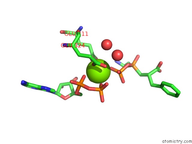
Mono view
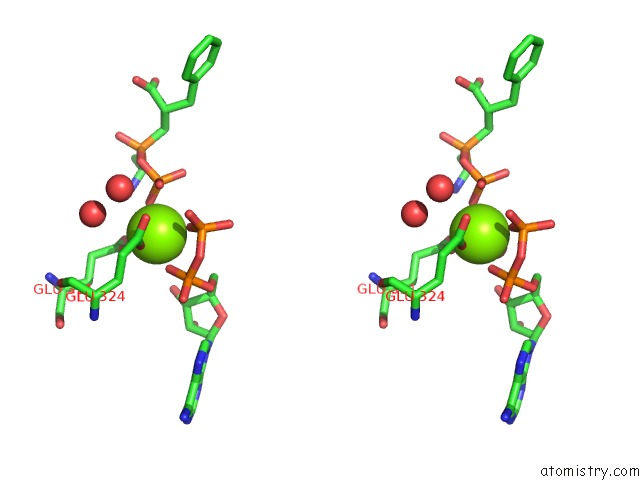
Stereo pair view
Mono view
Stereo pair view
A full contact list of Magnesium with other atoms in the Mg binding
site number 1 of Crystal Structure of Bacd, An L-Amino Acid Dipeptide Ligase From Bacillus Subtilis within 5.0Å range:
|
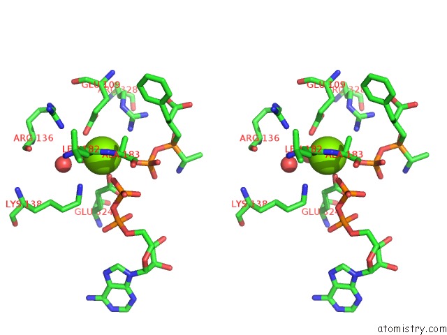
Magnesium binding site 2 out of 2 in 3vmm
Go back to
Magnesium binding site 2 out
of 2 in the Crystal Structure of Bacd, An L-Amino Acid Dipeptide Ligase From Bacillus Subtilis
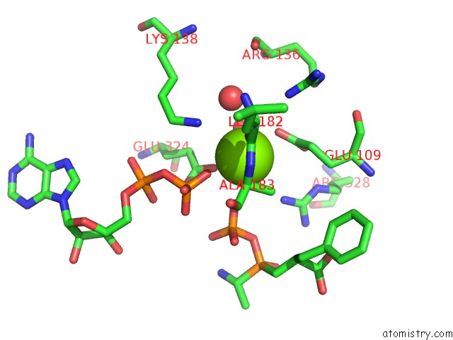
Mono view
Stereo pair view
Mono view
Stereo pair view
A full contact list of Magnesium with other atoms in the Mg binding
site number 2 of Crystal Structure of Bacd, An L-Amino Acid Dipeptide Ligase From Bacillus Subtilis within 5.0Å range:
|
Reference:
Y.Shomura,
E.Hinokuchi,
H.Ikeda,
A.Senoo,
Y.Takahashi,
J.Saito,
H.Komori,
N.Shibata,
Y.Yonetani,
Y.Higuchi.
Structural and Enzymatic Characterization of Bacd, An L-Amino Acid Dipeptide Ligase From Bacillus Subtilis Protein Sci. 2012.
ISSN: ESSN 1469-896X
PubMed: 22407814
DOI: 10.1002/PRO.2058
Page generated: Thu Aug 15 13:05:39 2024
ISSN: ESSN 1469-896X
PubMed: 22407814
DOI: 10.1002/PRO.2058
Last articles
Zn in 9J0NZn in 9J0O
Zn in 9J0P
Zn in 9FJX
Zn in 9EKB
Zn in 9C0F
Zn in 9CAH
Zn in 9CH0
Zn in 9CH3
Zn in 9CH1