Magnesium »
PDB 3vdb-3vsq »
3vmq »
Magnesium in PDB 3vmq: Crystal Structure of Staphylococcus Aureus Membrane-Bound Transglycosylase: Apoenzyme
Protein crystallography data
The structure of Crystal Structure of Staphylococcus Aureus Membrane-Bound Transglycosylase: Apoenzyme, PDB code: 3vmq
was solved by
C.Y.Huang,
H.W.Shih,
L.Y.Lin,
Y.W.Tien,
T.J.R.Cheng,
W.C.Cheng,
C.H.Wong,
C.Ma,
with X-Ray Crystallography technique. A brief refinement statistics is given in the table below:
| Resolution Low / High (Å) | 30.08 / 2.52 |
| Space group | P 21 21 21 |
| Cell size a, b, c (Å), α, β, γ (°) | 66.683, 67.406, 152.810, 90.00, 90.00, 90.00 |
| R / Rfree (%) | 20 / 25.7 |
Magnesium Binding Sites:
The binding sites of Magnesium atom in the Crystal Structure of Staphylococcus Aureus Membrane-Bound Transglycosylase: Apoenzyme
(pdb code 3vmq). This binding sites where shown within
5.0 Angstroms radius around Magnesium atom.
In total only one binding site of Magnesium was determined in the Crystal Structure of Staphylococcus Aureus Membrane-Bound Transglycosylase: Apoenzyme, PDB code: 3vmq:
In total only one binding site of Magnesium was determined in the Crystal Structure of Staphylococcus Aureus Membrane-Bound Transglycosylase: Apoenzyme, PDB code: 3vmq:
Magnesium binding site 1 out of 1 in 3vmq
Go back to
Magnesium binding site 1 out
of 1 in the Crystal Structure of Staphylococcus Aureus Membrane-Bound Transglycosylase: Apoenzyme
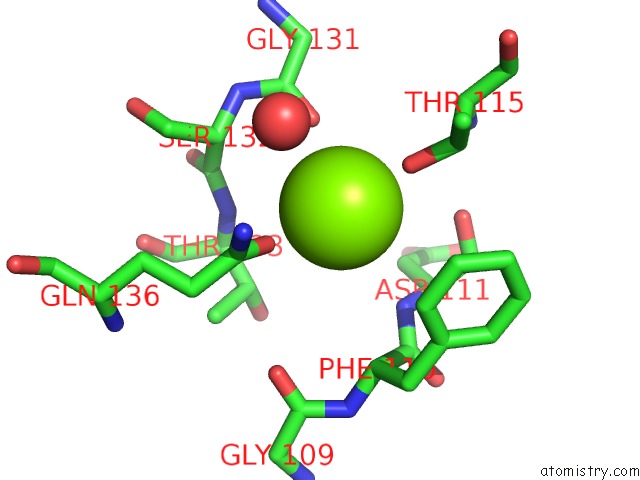
Mono view
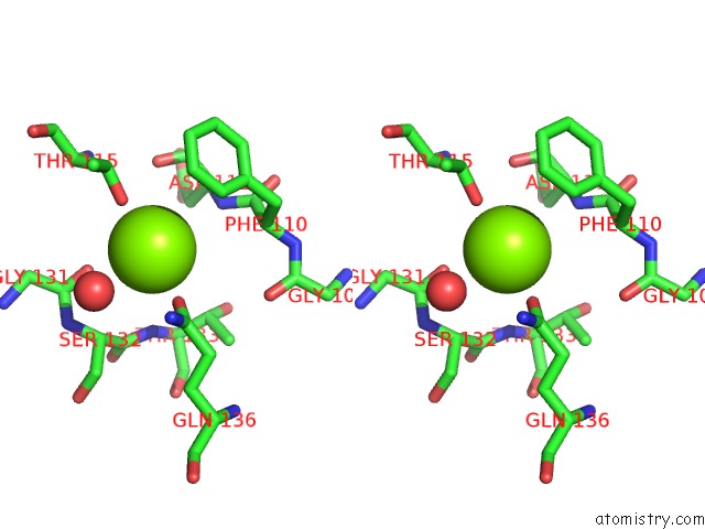
Stereo pair view
Mono view
Stereo pair view
A full contact list of Magnesium with other atoms in the Mg binding
site number 1 of Crystal Structure of Staphylococcus Aureus Membrane-Bound Transglycosylase: Apoenzyme within 5.0Å range:
|
Reference:
C.Y.Huang,
H.W.Shih,
L.Y.Lin,
Y.W.Tien,
T.J.R.Cheng,
W.C.Cheng,
C.H.Wong,
C.Ma.
Crystal Structure of Staphylococcus Aureus Transglycosylase in Complex with A Lipid II Analog and Elucidation of Peptidoglycan Synthesis Mechanism Proc.Natl.Acad.Sci.Usa V. 109 6496 2012.
ISSN: ISSN 0027-8424
PubMed: 22493270
DOI: 10.1073/PNAS.1203900109
Page generated: Thu Aug 15 13:05:46 2024
ISSN: ISSN 0027-8424
PubMed: 22493270
DOI: 10.1073/PNAS.1203900109
Last articles
Zn in 9J0NZn in 9J0O
Zn in 9J0P
Zn in 9FJX
Zn in 9EKB
Zn in 9C0F
Zn in 9CAH
Zn in 9CH0
Zn in 9CH3
Zn in 9CH1