Magnesium »
PDB 3vdc-3vth »
3vn9 »
Magnesium in PDB 3vn9: Rifined Crystal Structure of Non-Phosphorylated MAP2K6 in A Putative Auto-Inhibition State
Enzymatic activity of Rifined Crystal Structure of Non-Phosphorylated MAP2K6 in A Putative Auto-Inhibition State
All present enzymatic activity of Rifined Crystal Structure of Non-Phosphorylated MAP2K6 in A Putative Auto-Inhibition State:
2.7.12.2;
2.7.12.2;
Protein crystallography data
The structure of Rifined Crystal Structure of Non-Phosphorylated MAP2K6 in A Putative Auto-Inhibition State, PDB code: 3vn9
was solved by
T.Kinoshita,
H.Matsuzaka,
R.Nakai,
Y.Kirii,
K.Yokota,
T.Tada,
T.Matsumoto,
with X-Ray Crystallography technique. A brief refinement statistics is given in the table below:
| Resolution Low / High (Å) | 20.00 / 2.60 |
| Space group | P 31 2 1 |
| Cell size a, b, c (Å), α, β, γ (°) | 83.456, 83.456, 101.153, 90.00, 90.00, 120.00 |
| R / Rfree (%) | 26.4 / 28 |
Magnesium Binding Sites:
The binding sites of Magnesium atom in the Rifined Crystal Structure of Non-Phosphorylated MAP2K6 in A Putative Auto-Inhibition State
(pdb code 3vn9). This binding sites where shown within
5.0 Angstroms radius around Magnesium atom.
In total only one binding site of Magnesium was determined in the Rifined Crystal Structure of Non-Phosphorylated MAP2K6 in A Putative Auto-Inhibition State, PDB code: 3vn9:
In total only one binding site of Magnesium was determined in the Rifined Crystal Structure of Non-Phosphorylated MAP2K6 in A Putative Auto-Inhibition State, PDB code: 3vn9:
Magnesium binding site 1 out of 1 in 3vn9
Go back to
Magnesium binding site 1 out
of 1 in the Rifined Crystal Structure of Non-Phosphorylated MAP2K6 in A Putative Auto-Inhibition State
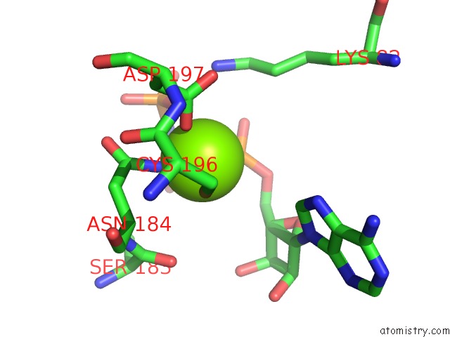
Mono view
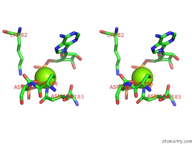
Stereo pair view
Mono view
Stereo pair view
A full contact list of Magnesium with other atoms in the Mg binding
site number 1 of Rifined Crystal Structure of Non-Phosphorylated MAP2K6 in A Putative Auto-Inhibition State within 5.0Å range:
|
Reference:
T.Matsumoto,
T.Kinoshita,
H.Matsuzaka,
R.Nakai,
Y.Kirii,
K.Yokota,
T.Tada.
Crystal Structure of Non-Phosphorylated MAP2K6 in A Putative Auto-Inhibition State J.Biochem. V. 151 541 2012.
ISSN: ISSN 0021-924X
PubMed: 22383536
DOI: 10.1093/JB/MVS023
Page generated: Mon Aug 11 04:39:42 2025
ISSN: ISSN 0021-924X
PubMed: 22383536
DOI: 10.1093/JB/MVS023
Last articles
Mg in 4EM4Mg in 4EMW
Mg in 4ELT
Mg in 4ELU
Mg in 4ELV
Mg in 4EM3
Mg in 4EKD
Mg in 4EHU
Mg in 4EHT
Mg in 4EKC