Magnesium »
PDB 3vdc-3vth »
3vsq »
Magnesium in PDB 3vsq: Crystal Structure of the Cytoplasmic Domain of G-Protein-Gated Inward Rectifier Potassium Channel KIR3.2 E236R Mutant in the Presence of Ethanol
Protein crystallography data
The structure of Crystal Structure of the Cytoplasmic Domain of G-Protein-Gated Inward Rectifier Potassium Channel KIR3.2 E236R Mutant in the Presence of Ethanol, PDB code: 3vsq
was solved by
A.Inanobe,
Y.Kurachi,
with X-Ray Crystallography technique. A brief refinement statistics is given in the table below:
| Resolution Low / High (Å) | 30.00 / 2.00 |
| Space group | P 4 21 2 |
| Cell size a, b, c (Å), α, β, γ (°) | 85.863, 85.863, 73.046, 90.00, 90.00, 90.00 |
| R / Rfree (%) | 21.6 / 25.9 |
Magnesium Binding Sites:
The binding sites of Magnesium atom in the Crystal Structure of the Cytoplasmic Domain of G-Protein-Gated Inward Rectifier Potassium Channel KIR3.2 E236R Mutant in the Presence of Ethanol
(pdb code 3vsq). This binding sites where shown within
5.0 Angstroms radius around Magnesium atom.
In total only one binding site of Magnesium was determined in the Crystal Structure of the Cytoplasmic Domain of G-Protein-Gated Inward Rectifier Potassium Channel KIR3.2 E236R Mutant in the Presence of Ethanol, PDB code: 3vsq:
In total only one binding site of Magnesium was determined in the Crystal Structure of the Cytoplasmic Domain of G-Protein-Gated Inward Rectifier Potassium Channel KIR3.2 E236R Mutant in the Presence of Ethanol, PDB code: 3vsq:
Magnesium binding site 1 out of 1 in 3vsq
Go back to
Magnesium binding site 1 out
of 1 in the Crystal Structure of the Cytoplasmic Domain of G-Protein-Gated Inward Rectifier Potassium Channel KIR3.2 E236R Mutant in the Presence of Ethanol
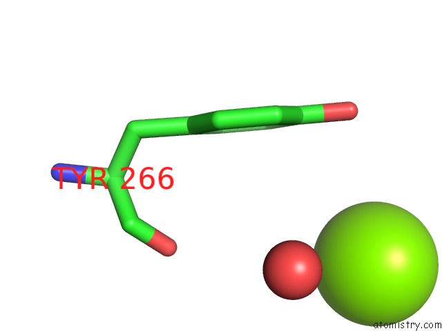
Mono view
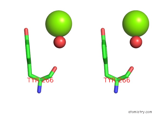
Stereo pair view
Mono view
Stereo pair view
A full contact list of Magnesium with other atoms in the Mg binding
site number 1 of Crystal Structure of the Cytoplasmic Domain of G-Protein-Gated Inward Rectifier Potassium Channel KIR3.2 E236R Mutant in the Presence of Ethanol within 5.0Å range:
|
Reference:
A.Inanobe,
Y.Kurachi.
Coupling of G Protein Binding to Channel Gating in Mammalian Inward Rectifier K+ Channels To Be Published.
Page generated: Mon Aug 11 04:41:01 2025
Last articles
Mg in 4UM5Mg in 4UKD
Mg in 4UJ3
Mg in 4UJ5
Mg in 4UJ4
Mg in 4UHS
Mg in 4UHO
Mg in 4UHN
Mg in 4UHM
Mg in 4UHK