Magnesium »
PDB 3wdl-3wqc »
3wg7 »
Magnesium in PDB 3wg7: A 1.9 Angstrom Radiation Damage Free X-Ray Structure of Large (420KDA) Protein By Femtosecond Crystallography
Enzymatic activity of A 1.9 Angstrom Radiation Damage Free X-Ray Structure of Large (420KDA) Protein By Femtosecond Crystallography
All present enzymatic activity of A 1.9 Angstrom Radiation Damage Free X-Ray Structure of Large (420KDA) Protein By Femtosecond Crystallography:
1.9.3.1;
1.9.3.1;
Protein crystallography data
The structure of A 1.9 Angstrom Radiation Damage Free X-Ray Structure of Large (420KDA) Protein By Femtosecond Crystallography, PDB code: 3wg7
was solved by
K.Hirata,
K.Shinzawa-Itoh,
N.Yano,
S.Takemura,
K.Kato,
M.Hatanaka,
K.Muramoto,
T.Kawahara,
T.Tsukihara,
E.Yamashita,
K.Tono,
G.Ueno,
T.Hikima,
H.Murakami,
Y.Inubushi,
M.Yabashi,
T.Ishikawa,
M.Yamamoto,
T.Ogura,
H.Sugimoto,
J.R.Shen,
S.Yoshikawa,
H.Ago,
with X-Ray Crystallography technique. A brief refinement statistics is given in the table below:
| Resolution Low / High (Å) | 40.00 / 1.90 |
| Space group | P 21 21 21 |
| Cell size a, b, c (Å), α, β, γ (°) | 182.600, 204.510, 178.290, 90.00, 90.00, 90.00 |
| R / Rfree (%) | 19.5 / 23 |
Other elements in 3wg7:
The structure of A 1.9 Angstrom Radiation Damage Free X-Ray Structure of Large (420KDA) Protein By Femtosecond Crystallography also contains other interesting chemical elements:
| Zinc | (Zn) | 2 atoms |
| Iron | (Fe) | 4 atoms |
| Copper | (Cu) | 6 atoms |
| Sodium | (Na) | 4 atoms |
Magnesium Binding Sites:
The binding sites of Magnesium atom in the A 1.9 Angstrom Radiation Damage Free X-Ray Structure of Large (420KDA) Protein By Femtosecond Crystallography
(pdb code 3wg7). This binding sites where shown within
5.0 Angstroms radius around Magnesium atom.
In total 2 binding sites of Magnesium where determined in the A 1.9 Angstrom Radiation Damage Free X-Ray Structure of Large (420KDA) Protein By Femtosecond Crystallography, PDB code: 3wg7:
Jump to Magnesium binding site number: 1; 2;
In total 2 binding sites of Magnesium where determined in the A 1.9 Angstrom Radiation Damage Free X-Ray Structure of Large (420KDA) Protein By Femtosecond Crystallography, PDB code: 3wg7:
Jump to Magnesium binding site number: 1; 2;
Magnesium binding site 1 out of 2 in 3wg7
Go back to
Magnesium binding site 1 out
of 2 in the A 1.9 Angstrom Radiation Damage Free X-Ray Structure of Large (420KDA) Protein By Femtosecond Crystallography
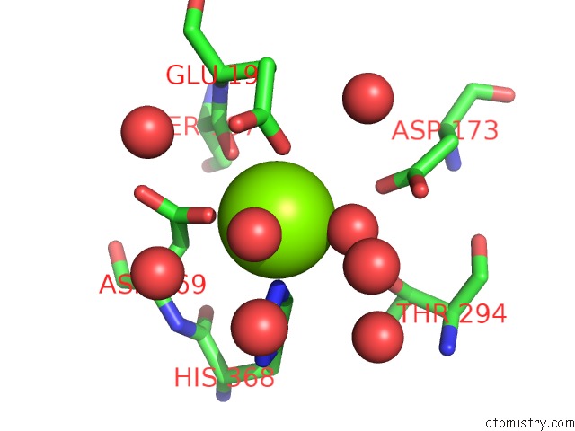
Mono view
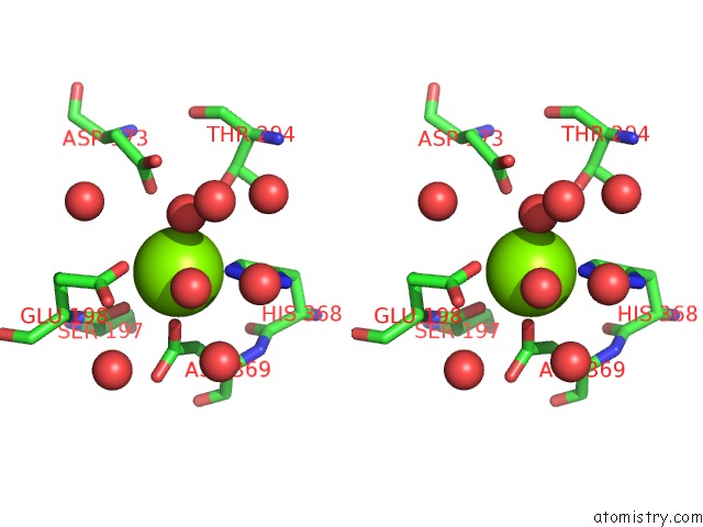
Stereo pair view
Mono view
Stereo pair view
A full contact list of Magnesium with other atoms in the Mg binding
site number 1 of A 1.9 Angstrom Radiation Damage Free X-Ray Structure of Large (420KDA) Protein By Femtosecond Crystallography within 5.0Å range:
|
Magnesium binding site 2 out of 2 in 3wg7
Go back to
Magnesium binding site 2 out
of 2 in the A 1.9 Angstrom Radiation Damage Free X-Ray Structure of Large (420KDA) Protein By Femtosecond Crystallography
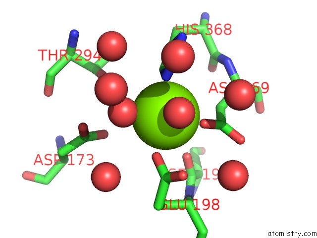
Mono view
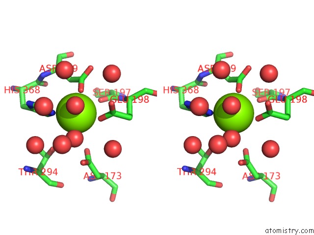
Stereo pair view
Mono view
Stereo pair view
A full contact list of Magnesium with other atoms in the Mg binding
site number 2 of A 1.9 Angstrom Radiation Damage Free X-Ray Structure of Large (420KDA) Protein By Femtosecond Crystallography within 5.0Å range:
|
Reference:
K.Hirata,
K.Shinzawa-Itoh,
N.Yano,
S.Takemura,
K.Kato,
M.Hatanaka,
K.Muramoto,
T.Kawahara,
T.Tsukihara,
E.Yamashita,
K.Tono,
G.Ueno,
T.Hikima,
H.Murakami,
Y.Inubushi,
M.Yabashi,
T.Ishikawa,
M.Yamamoto,
T.Ogura,
H.Sugimoto,
J.R.Shen,
S.Yoshikawa,
H.Ago.
Determination of Damage-Free Crystal Structure of An X-Ray-Sensitive Protein Using An Xfel. Nat.Methods 2014.
ISSN: ESSN 1548-7105
PubMed: 24813624
DOI: 10.1038/NMETH.2962
Page generated: Thu Aug 15 13:24:13 2024
ISSN: ESSN 1548-7105
PubMed: 24813624
DOI: 10.1038/NMETH.2962
Last articles
Zn in 9MJ5Zn in 9HNW
Zn in 9G0L
Zn in 9FNE
Zn in 9DZN
Zn in 9E0I
Zn in 9D32
Zn in 9DAK
Zn in 8ZXC
Zn in 8ZUF