Magnesium »
PDB 3wcw-3wow »
3wk6 »
Magnesium in PDB 3wk6: Crystal Structure of Soluble Epoxide Hydrolase in Complex with Fragment Inhibitor
Enzymatic activity of Crystal Structure of Soluble Epoxide Hydrolase in Complex with Fragment Inhibitor
All present enzymatic activity of Crystal Structure of Soluble Epoxide Hydrolase in Complex with Fragment Inhibitor:
3.1.3.76; 3.3.2.10;
3.1.3.76; 3.3.2.10;
Protein crystallography data
The structure of Crystal Structure of Soluble Epoxide Hydrolase in Complex with Fragment Inhibitor, PDB code: 3wk6
was solved by
Y.Amano,
T.Yamaguchi,
E.Tanabe,
with X-Ray Crystallography technique. A brief refinement statistics is given in the table below:
| Resolution Low / High (Å) | 46.48 / 2.10 |
| Space group | P 65 2 2 |
| Cell size a, b, c (Å), α, β, γ (°) | 92.952, 92.952, 243.795, 90.00, 90.00, 120.00 |
| R / Rfree (%) | 19.6 / 25 |
Magnesium Binding Sites:
The binding sites of Magnesium atom in the Crystal Structure of Soluble Epoxide Hydrolase in Complex with Fragment Inhibitor
(pdb code 3wk6). This binding sites where shown within
5.0 Angstroms radius around Magnesium atom.
In total only one binding site of Magnesium was determined in the Crystal Structure of Soluble Epoxide Hydrolase in Complex with Fragment Inhibitor, PDB code: 3wk6:
In total only one binding site of Magnesium was determined in the Crystal Structure of Soluble Epoxide Hydrolase in Complex with Fragment Inhibitor, PDB code: 3wk6:
Magnesium binding site 1 out of 1 in 3wk6
Go back to
Magnesium binding site 1 out
of 1 in the Crystal Structure of Soluble Epoxide Hydrolase in Complex with Fragment Inhibitor
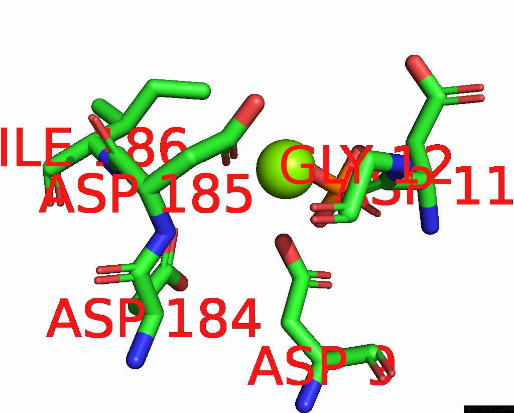
Mono view
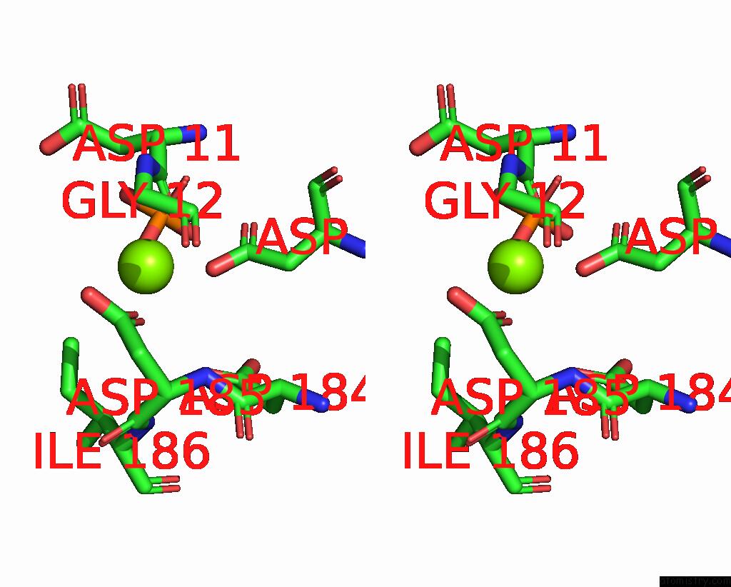
Stereo pair view
Mono view
Stereo pair view
A full contact list of Magnesium with other atoms in the Mg binding
site number 1 of Crystal Structure of Soluble Epoxide Hydrolase in Complex with Fragment Inhibitor within 5.0Å range:
|
Reference:
Y.Amano,
T.Yamaguchi,
E.Tanabe.
Structural Insights Into Binding of Inhibitors to Soluble Epoxide Hydrolase Gained By Fragment Screening and X-Ray Crystallography Bioorg.Med.Chem. 2014.
ISSN: ESSN 1464-3391
PubMed: 24656800
DOI: 10.1016/J.BMC.2014.03.001
Page generated: Thu Aug 15 13:26:06 2024
ISSN: ESSN 1464-3391
PubMed: 24656800
DOI: 10.1016/J.BMC.2014.03.001
Last articles
Zn in 9J0NZn in 9J0O
Zn in 9J0P
Zn in 9FJX
Zn in 9EKB
Zn in 9C0F
Zn in 9CAH
Zn in 9CH0
Zn in 9CH3
Zn in 9CH1