Magnesium »
PDB 3wqc-3x1d »
3wqm »
Magnesium in PDB 3wqm: Crystal Structure of RV3378C with Inhibitor Bph-629
Enzymatic activity of Crystal Structure of RV3378C with Inhibitor Bph-629
All present enzymatic activity of Crystal Structure of RV3378C with Inhibitor Bph-629:
3.1.7.8; 3.1.7.9;
3.1.7.8; 3.1.7.9;
Protein crystallography data
The structure of Crystal Structure of RV3378C with Inhibitor Bph-629, PDB code: 3wqm
was solved by
H.C.Chan,
X.Feng,
T.P.Ko,
C.H.Huang,
Y.Hu,
Y.Zheng,
S.Bogue,
C.Nakano,
T.Hoshino,
L.Zhang,
P.Lv,
W.Liu,
D.C.Crick,
P.H.Liang,
A.H.Wang,
E.Oldfield,
R.T.Guo,
with X-Ray Crystallography technique. A brief refinement statistics is given in the table below:
| Resolution Low / High (Å) | 25.00 / 2.10 |
| Space group | P 43 21 2 |
| Cell size a, b, c (Å), α, β, γ (°) | 105.417, 105.417, 65.991, 90.00, 90.00, 90.00 |
| R / Rfree (%) | 19.7 / 23.7 |
Magnesium Binding Sites:
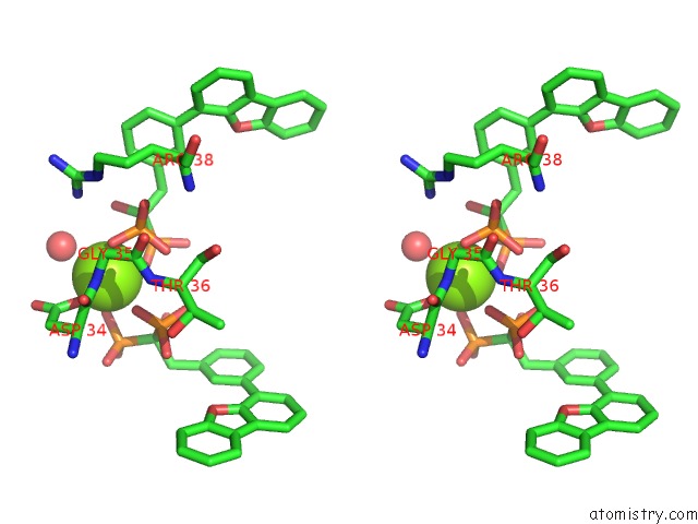
The binding sites of Magnesium atom in the Crystal Structure of RV3378C with Inhibitor Bph-629
(pdb code 3wqm). This binding sites where shown within
5.0 Angstroms radius around Magnesium atom.
In total only one binding site of Magnesium was determined in the Crystal Structure of RV3378C with Inhibitor Bph-629, PDB code: 3wqm:
In total only one binding site of Magnesium was determined in the Crystal Structure of RV3378C with Inhibitor Bph-629, PDB code: 3wqm:
Magnesium binding site 1 out of 1 in 3wqm
Go back to
Magnesium binding site 1 out
of 1 in the Crystal Structure of RV3378C with Inhibitor Bph-629

Mono view
Stereo pair view
Mono view
Stereo pair view
A full contact list of Magnesium with other atoms in the Mg binding
site number 1 of Crystal Structure of RV3378C with Inhibitor Bph-629 within 5.0Å range:
|
Reference:
H.C.Chan,
X.Feng,
T.P.Ko,
C.H.Huang,
Y.Hu,
Y.Zheng,
S.Bogue,
C.Nakano,
T.Hoshino,
L.Zhang,
P.Lv,
W.Liu,
D.C.Crick,
P.H.Liang,
A.H.Wang,
E.Oldfield,
R.T.Guo.
Structure and Inhibition of Tuberculosinol Synthase and Decaprenyl Diphosphate Synthase From Mycobacterium Tuberculosis. J.Am.Chem.Soc. V. 136 2892 2014.
ISSN: ISSN 0002-7863
PubMed: 24475925
DOI: 10.1021/JA413127V
Page generated: Thu Aug 15 13:33:21 2024
ISSN: ISSN 0002-7863
PubMed: 24475925
DOI: 10.1021/JA413127V
Last articles
Zn in 9J0NZn in 9J0O
Zn in 9J0P
Zn in 9FJX
Zn in 9EKB
Zn in 9C0F
Zn in 9CAH
Zn in 9CH0
Zn in 9CH3
Zn in 9CH1