Magnesium »
PDB 3wqd-3x1l »
3wqp »
Magnesium in PDB 3wqp: Crystal Structure of Rubisco T289D Mutant From Thermococcus Kodakarensis
Enzymatic activity of Crystal Structure of Rubisco T289D Mutant From Thermococcus Kodakarensis
All present enzymatic activity of Crystal Structure of Rubisco T289D Mutant From Thermococcus Kodakarensis:
4.1.1.39;
4.1.1.39;
Protein crystallography data
The structure of Crystal Structure of Rubisco T289D Mutant From Thermococcus Kodakarensis, PDB code: 3wqp
was solved by
M.Fujihashi,
Y.Nishitani,
T.Kiriyama,
K.Miki,
with X-Ray Crystallography technique. A brief refinement statistics is given in the table below:
| Resolution Low / High (Å) | 50.00 / 2.25 |
| Space group | P 21 21 2 |
| Cell size a, b, c (Å), α, β, γ (°) | 170.566, 246.238, 144.628, 90.00, 90.00, 90.00 |
| R / Rfree (%) | 22 / 25.8 |
Magnesium Binding Sites:
The binding sites of Magnesium atom in the Crystal Structure of Rubisco T289D Mutant From Thermococcus Kodakarensis
(pdb code 3wqp). This binding sites where shown within
5.0 Angstroms radius around Magnesium atom.
In total 10 binding sites of Magnesium where determined in the Crystal Structure of Rubisco T289D Mutant From Thermococcus Kodakarensis, PDB code: 3wqp:
Jump to Magnesium binding site number: 1; 2; 3; 4; 5; 6; 7; 8; 9; 10;
In total 10 binding sites of Magnesium where determined in the Crystal Structure of Rubisco T289D Mutant From Thermococcus Kodakarensis, PDB code: 3wqp:
Jump to Magnesium binding site number: 1; 2; 3; 4; 5; 6; 7; 8; 9; 10;
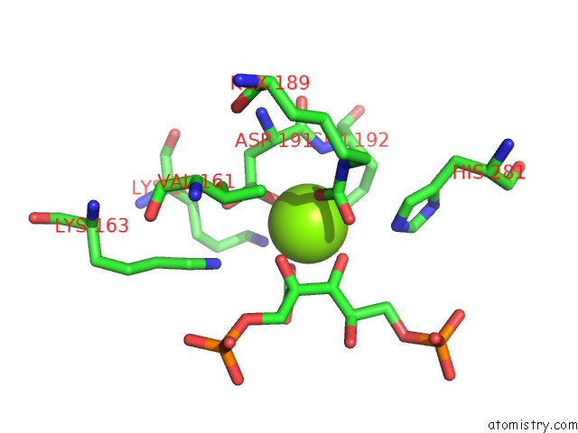
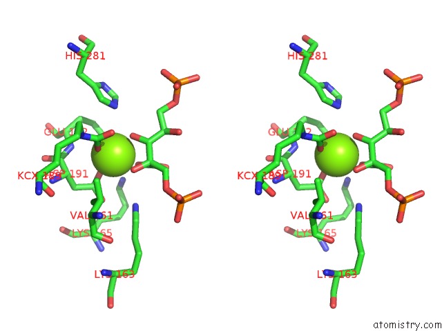
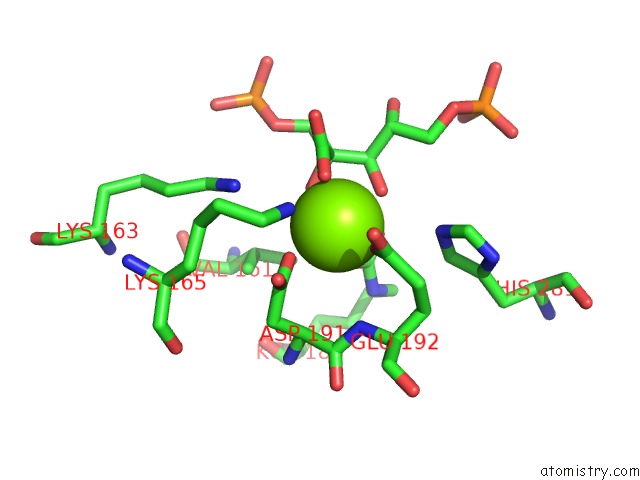
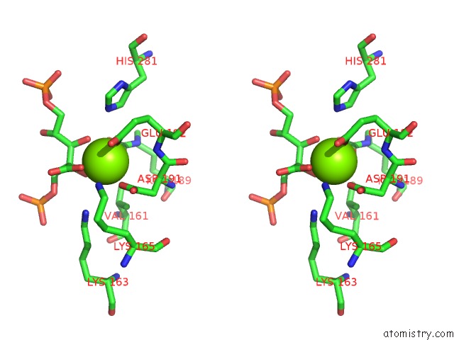
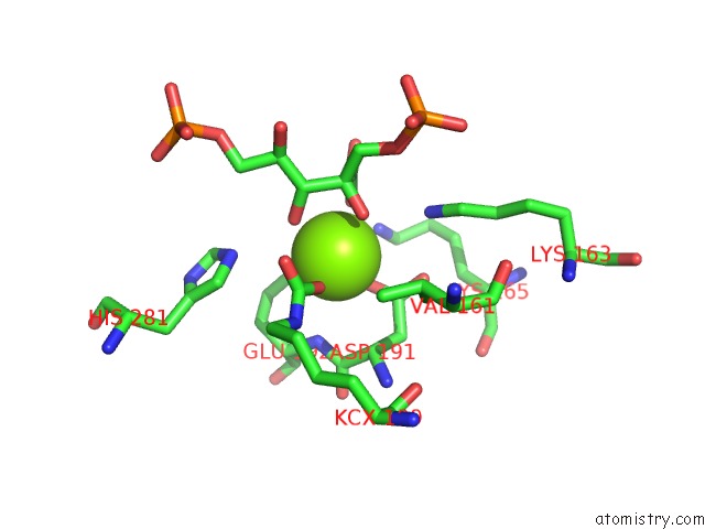
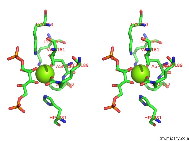
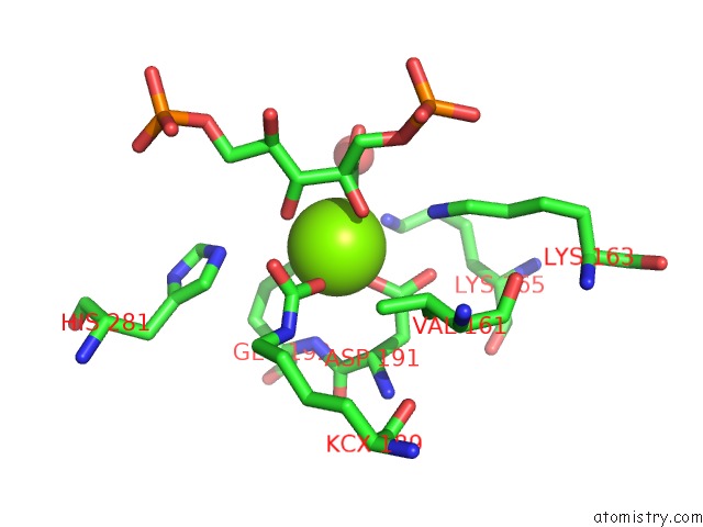
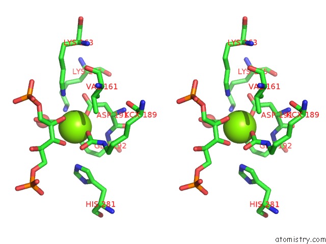
Magnesium binding site 1 out of 10 in 3wqp
Go back to
Magnesium binding site 1 out
of 10 in the Crystal Structure of Rubisco T289D Mutant From Thermococcus Kodakarensis
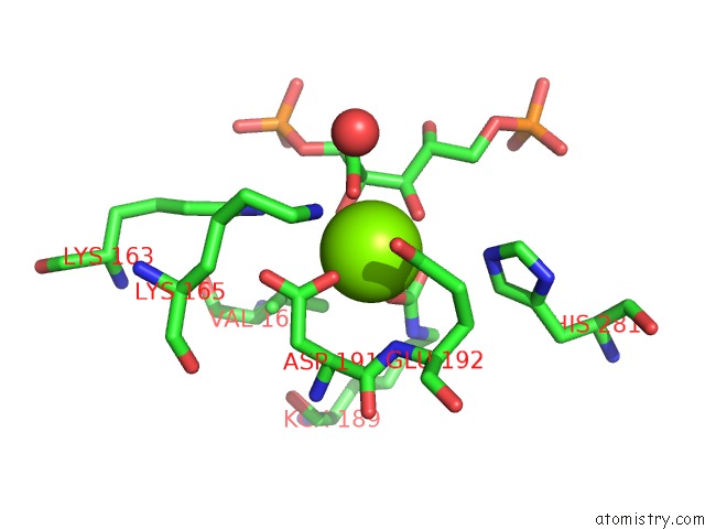
Mono view
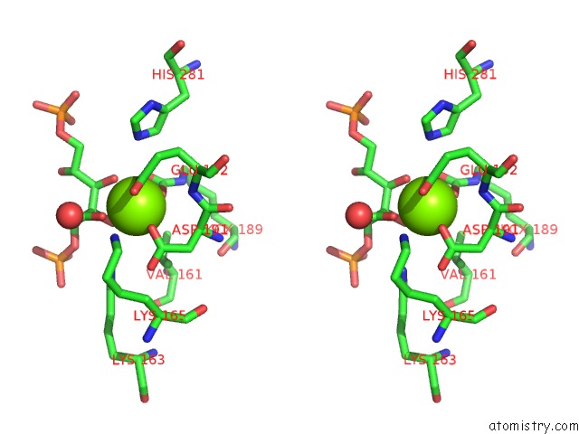
Stereo pair view
Mono view
Stereo pair view
A full contact list of Magnesium with other atoms in the Mg binding
site number 1 of Crystal Structure of Rubisco T289D Mutant From Thermococcus Kodakarensis within 5.0Å range:
|
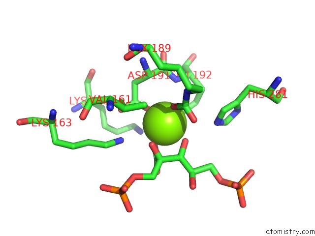
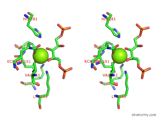
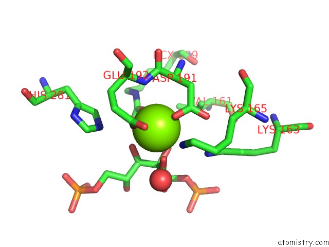
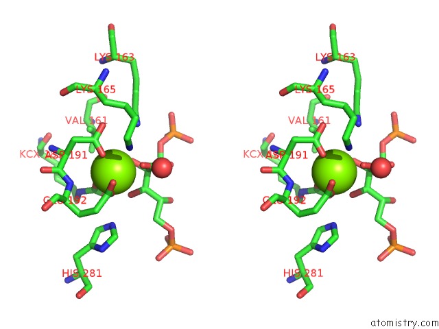
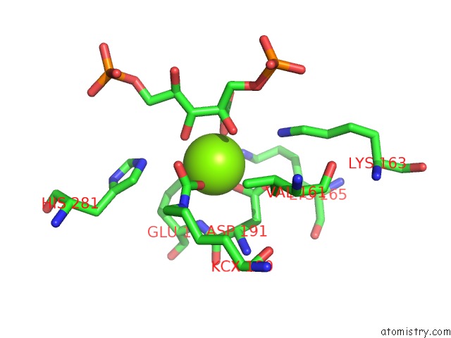
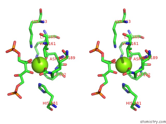
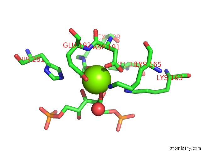
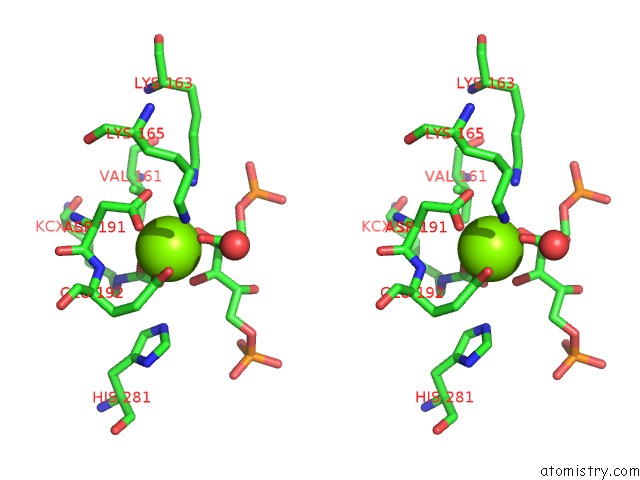
Magnesium binding site 2 out of 10 in 3wqp
Go back to
Magnesium binding site 2 out
of 10 in the Crystal Structure of Rubisco T289D Mutant From Thermococcus Kodakarensis
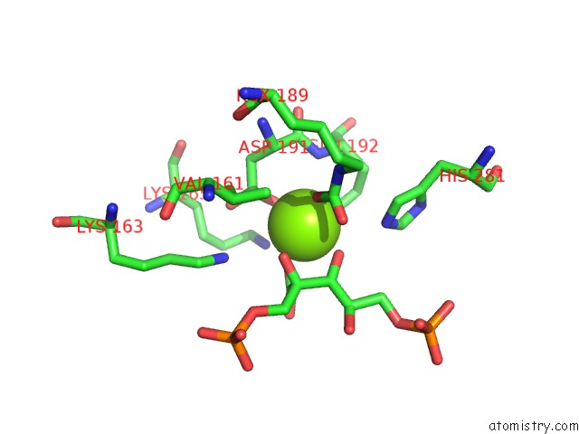
Mono view
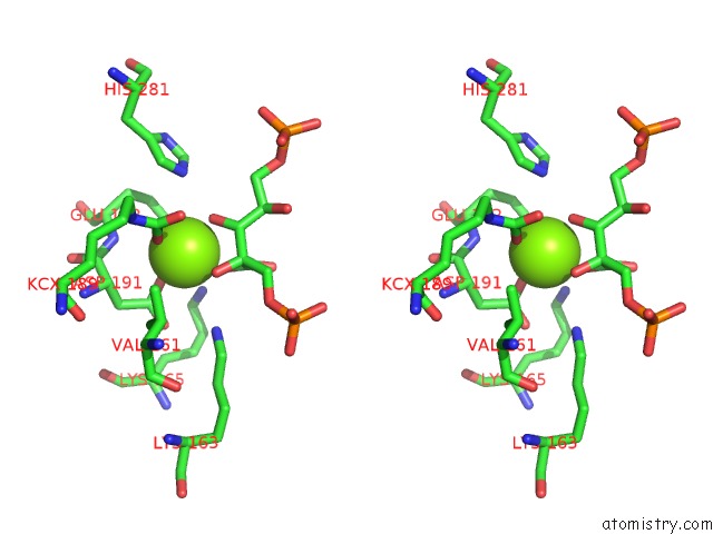
Stereo pair view
Mono view
Stereo pair view
A full contact list of Magnesium with other atoms in the Mg binding
site number 2 of Crystal Structure of Rubisco T289D Mutant From Thermococcus Kodakarensis within 5.0Å range:
|
Magnesium binding site 3 out of 10 in 3wqp
Go back to
Magnesium binding site 3 out
of 10 in the Crystal Structure of Rubisco T289D Mutant From Thermococcus Kodakarensis
Mono view
Stereo pair view
Mono view
Stereo pair view
A full contact list of Magnesium with other atoms in the Mg binding
site number 3 of Crystal Structure of Rubisco T289D Mutant From Thermococcus Kodakarensis within 5.0Å range:
|
Magnesium binding site 4 out of 10 in 3wqp
Go back to
Magnesium binding site 4 out
of 10 in the Crystal Structure of Rubisco T289D Mutant From Thermococcus Kodakarensis
Mono view
Stereo pair view
Mono view
Stereo pair view
A full contact list of Magnesium with other atoms in the Mg binding
site number 4 of Crystal Structure of Rubisco T289D Mutant From Thermococcus Kodakarensis within 5.0Å range:
|
Magnesium binding site 5 out of 10 in 3wqp
Go back to
Magnesium binding site 5 out
of 10 in the Crystal Structure of Rubisco T289D Mutant From Thermococcus Kodakarensis
Mono view
Stereo pair view
Mono view
Stereo pair view
A full contact list of Magnesium with other atoms in the Mg binding
site number 5 of Crystal Structure of Rubisco T289D Mutant From Thermococcus Kodakarensis within 5.0Å range:
|
Magnesium binding site 6 out of 10 in 3wqp
Go back to
Magnesium binding site 6 out
of 10 in the Crystal Structure of Rubisco T289D Mutant From Thermococcus Kodakarensis
Mono view
Stereo pair view
Mono view
Stereo pair view
A full contact list of Magnesium with other atoms in the Mg binding
site number 6 of Crystal Structure of Rubisco T289D Mutant From Thermococcus Kodakarensis within 5.0Å range:
|
Magnesium binding site 7 out of 10 in 3wqp
Go back to
Magnesium binding site 7 out
of 10 in the Crystal Structure of Rubisco T289D Mutant From Thermococcus Kodakarensis
Mono view
Stereo pair view
Mono view
Stereo pair view
A full contact list of Magnesium with other atoms in the Mg binding
site number 7 of Crystal Structure of Rubisco T289D Mutant From Thermococcus Kodakarensis within 5.0Å range:
|
Magnesium binding site 8 out of 10 in 3wqp
Go back to
Magnesium binding site 8 out
of 10 in the Crystal Structure of Rubisco T289D Mutant From Thermococcus Kodakarensis
Mono view
Stereo pair view
Mono view
Stereo pair view
A full contact list of Magnesium with other atoms in the Mg binding
site number 8 of Crystal Structure of Rubisco T289D Mutant From Thermococcus Kodakarensis within 5.0Å range:
|
Magnesium binding site 9 out of 10 in 3wqp
Go back to
Magnesium binding site 9 out
of 10 in the Crystal Structure of Rubisco T289D Mutant From Thermococcus Kodakarensis
Mono view
Stereo pair view
Mono view
Stereo pair view
A full contact list of Magnesium with other atoms in the Mg binding
site number 9 of Crystal Structure of Rubisco T289D Mutant From Thermococcus Kodakarensis within 5.0Å range:
|
Magnesium binding site 10 out of 10 in 3wqp
Go back to
Magnesium binding site 10 out
of 10 in the Crystal Structure of Rubisco T289D Mutant From Thermococcus Kodakarensis
Mono view
Stereo pair view
Mono view
Stereo pair view
A full contact list of Magnesium with other atoms in the Mg binding
site number 10 of Crystal Structure of Rubisco T289D Mutant From Thermococcus Kodakarensis within 5.0Å range:
|
Reference:
T.Kiriyama,
M.Fujihashi,
Y.Nishitani,
R.Aono,
T.Sato,
T.Takai,
K.Tagashira,
W.Fukuda,
H.Atomi,
T.Imanaka,
K.Miki.
Mutation Design of Thermophilic Rubisco Based on the Three-Dimensional Structure Enhances Its Activity at Ambient Temperature To Be Published.
Page generated: Thu Aug 15 13:33:23 2024
Last articles
Fe in 2YXOFe in 2YRS
Fe in 2YXC
Fe in 2YNM
Fe in 2YVJ
Fe in 2YP1
Fe in 2YU2
Fe in 2YU1
Fe in 2YQB
Fe in 2YOO