Magnesium »
PDB 3wqd-3x1l »
3wvl »
Magnesium in PDB 3wvl: Crystal Structure of the Football-Shaped Groel-Groes Complex (Groel: GROES2:ATP14) From Escherichia Coli
Enzymatic activity of Crystal Structure of the Football-Shaped Groel-Groes Complex (Groel: GROES2:ATP14) From Escherichia Coli
All present enzymatic activity of Crystal Structure of the Football-Shaped Groel-Groes Complex (Groel: GROES2:ATP14) From Escherichia Coli:
3.6.4.9;
3.6.4.9;
Protein crystallography data
The structure of Crystal Structure of the Football-Shaped Groel-Groes Complex (Groel: GROES2:ATP14) From Escherichia Coli, PDB code: 3wvl
was solved by
A.Koike-Takeshita,
T.Arakawa,
H.Taguchi,
T.Shimamura,
with X-Ray Crystallography technique. A brief refinement statistics is given in the table below:
| Resolution Low / High (Å) | 49.56 / 3.79 |
| Space group | P 1 21 1 |
| Cell size a, b, c (Å), α, β, γ (°) | 160.280, 217.117, 198.481, 90.00, 98.02, 90.00 |
| R / Rfree (%) | 21.1 / 25 |
Other elements in 3wvl:
The structure of Crystal Structure of the Football-Shaped Groel-Groes Complex (Groel: GROES2:ATP14) From Escherichia Coli also contains other interesting chemical elements:
| Potassium | (K) | 14 atoms |
Magnesium Binding Sites:
Pages:
>>> Page 1 <<< Page 2, Binding sites: 11 - 14;Binding sites:
The binding sites of Magnesium atom in the Crystal Structure of the Football-Shaped Groel-Groes Complex (Groel: GROES2:ATP14) From Escherichia Coli (pdb code 3wvl). This binding sites where shown within 5.0 Angstroms radius around Magnesium atom.In total 14 binding sites of Magnesium where determined in the Crystal Structure of the Football-Shaped Groel-Groes Complex (Groel: GROES2:ATP14) From Escherichia Coli, PDB code: 3wvl:
Jump to Magnesium binding site number: 1; 2; 3; 4; 5; 6; 7; 8; 9; 10;
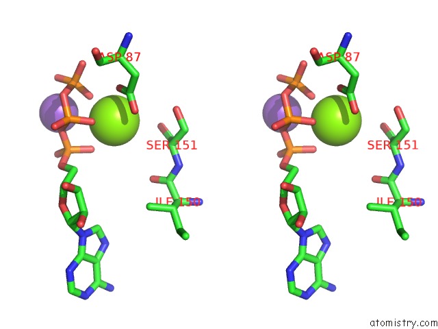
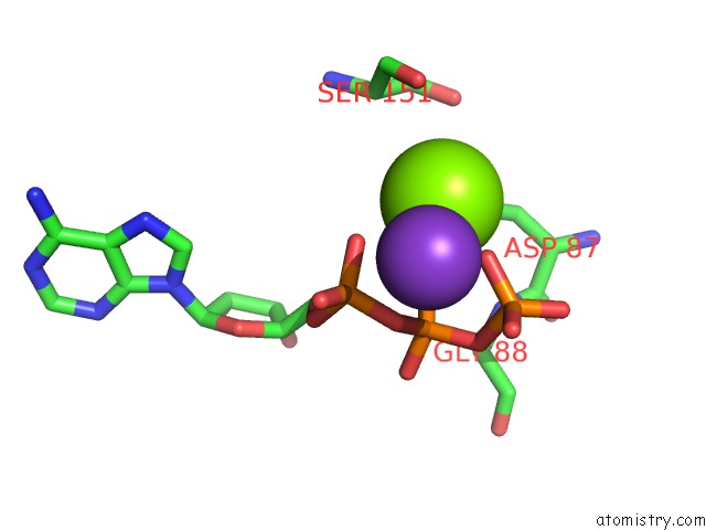
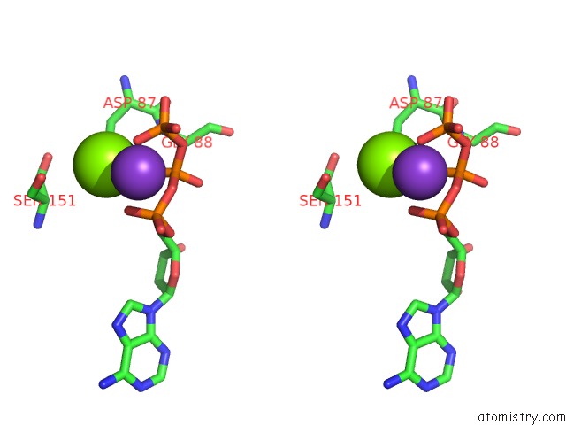
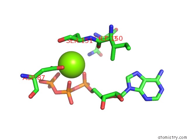
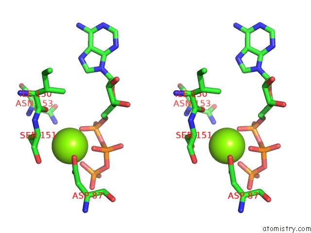
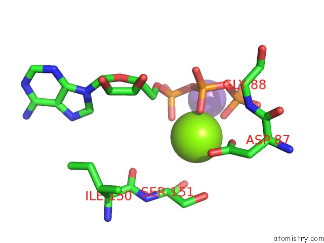
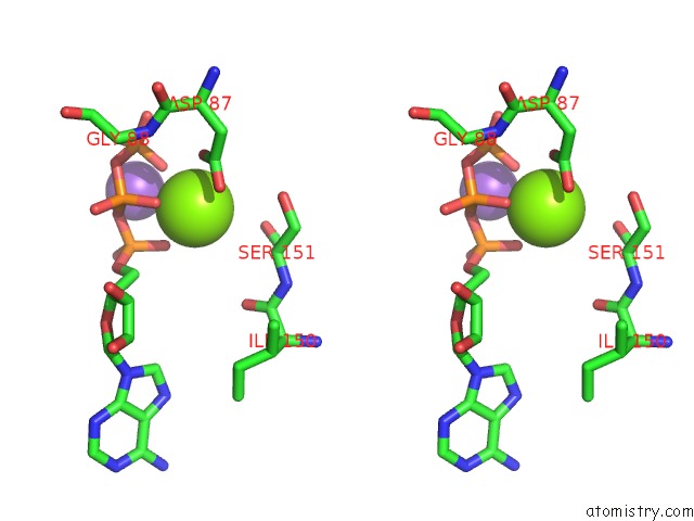
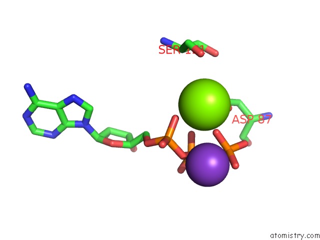
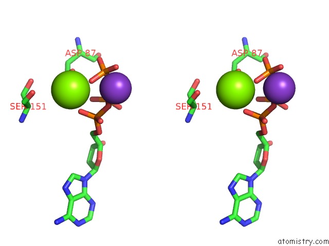
Magnesium binding site 1 out of 14 in 3wvl
Go back to
Magnesium binding site 1 out
of 14 in the Crystal Structure of the Football-Shaped Groel-Groes Complex (Groel: GROES2:ATP14) From Escherichia Coli
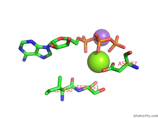
Mono view
Stereo pair view
Mono view
Stereo pair view
A full contact list of Magnesium with other atoms in the Mg binding
site number 1 of Crystal Structure of the Football-Shaped Groel-Groes Complex (Groel: GROES2:ATP14) From Escherichia Coli within 5.0Å range:
|
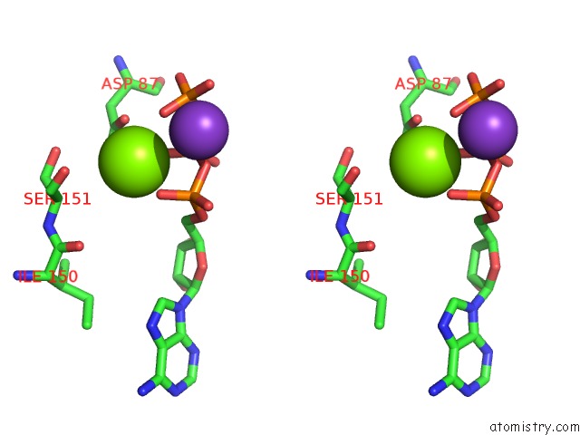
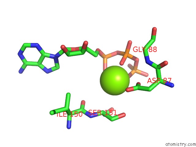
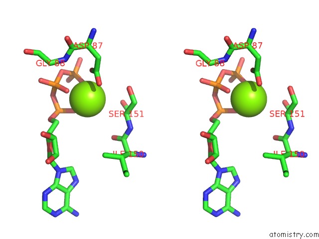
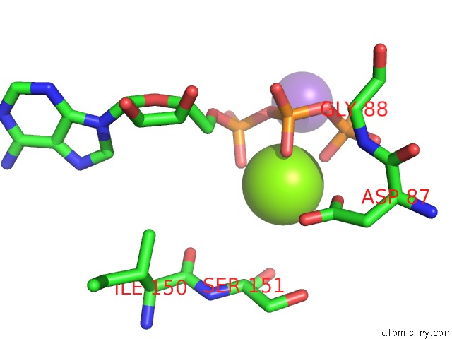
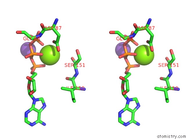
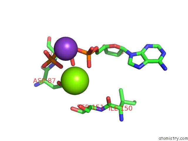
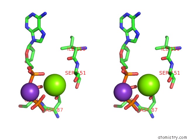
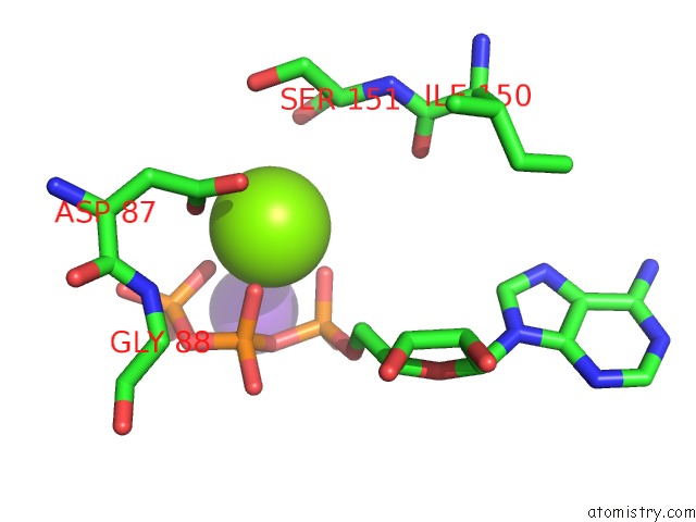
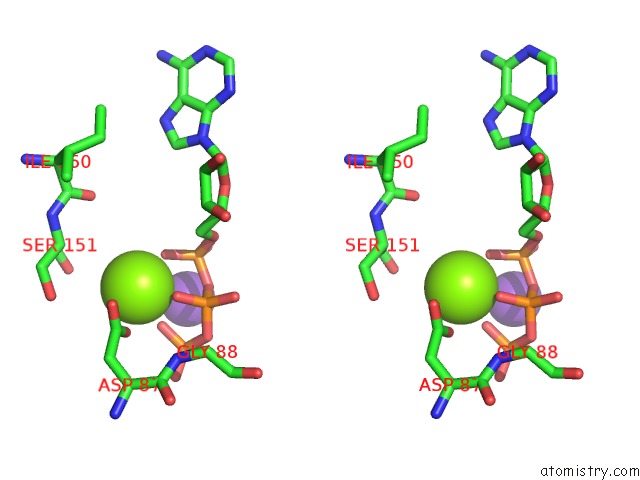
Magnesium binding site 2 out of 14 in 3wvl
Go back to
Magnesium binding site 2 out
of 14 in the Crystal Structure of the Football-Shaped Groel-Groes Complex (Groel: GROES2:ATP14) From Escherichia Coli
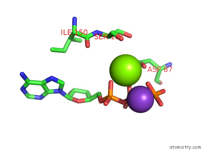
Mono view
Stereo pair view
Mono view
Stereo pair view
A full contact list of Magnesium with other atoms in the Mg binding
site number 2 of Crystal Structure of the Football-Shaped Groel-Groes Complex (Groel: GROES2:ATP14) From Escherichia Coli within 5.0Å range:
|
Magnesium binding site 3 out of 14 in 3wvl
Go back to
Magnesium binding site 3 out
of 14 in the Crystal Structure of the Football-Shaped Groel-Groes Complex (Groel: GROES2:ATP14) From Escherichia Coli
Mono view
Stereo pair view
Mono view
Stereo pair view
A full contact list of Magnesium with other atoms in the Mg binding
site number 3 of Crystal Structure of the Football-Shaped Groel-Groes Complex (Groel: GROES2:ATP14) From Escherichia Coli within 5.0Å range:
|
Magnesium binding site 4 out of 14 in 3wvl
Go back to
Magnesium binding site 4 out
of 14 in the Crystal Structure of the Football-Shaped Groel-Groes Complex (Groel: GROES2:ATP14) From Escherichia Coli
Mono view
Stereo pair view
Mono view
Stereo pair view
A full contact list of Magnesium with other atoms in the Mg binding
site number 4 of Crystal Structure of the Football-Shaped Groel-Groes Complex (Groel: GROES2:ATP14) From Escherichia Coli within 5.0Å range:
|
Magnesium binding site 5 out of 14 in 3wvl
Go back to
Magnesium binding site 5 out
of 14 in the Crystal Structure of the Football-Shaped Groel-Groes Complex (Groel: GROES2:ATP14) From Escherichia Coli
Mono view
Stereo pair view
Mono view
Stereo pair view
A full contact list of Magnesium with other atoms in the Mg binding
site number 5 of Crystal Structure of the Football-Shaped Groel-Groes Complex (Groel: GROES2:ATP14) From Escherichia Coli within 5.0Å range:
|
Magnesium binding site 6 out of 14 in 3wvl
Go back to
Magnesium binding site 6 out
of 14 in the Crystal Structure of the Football-Shaped Groel-Groes Complex (Groel: GROES2:ATP14) From Escherichia Coli
Mono view
Stereo pair view
Mono view
Stereo pair view
A full contact list of Magnesium with other atoms in the Mg binding
site number 6 of Crystal Structure of the Football-Shaped Groel-Groes Complex (Groel: GROES2:ATP14) From Escherichia Coli within 5.0Å range:
|
Magnesium binding site 7 out of 14 in 3wvl
Go back to
Magnesium binding site 7 out
of 14 in the Crystal Structure of the Football-Shaped Groel-Groes Complex (Groel: GROES2:ATP14) From Escherichia Coli
Mono view
Stereo pair view
Mono view
Stereo pair view
A full contact list of Magnesium with other atoms in the Mg binding
site number 7 of Crystal Structure of the Football-Shaped Groel-Groes Complex (Groel: GROES2:ATP14) From Escherichia Coli within 5.0Å range:
|
Magnesium binding site 8 out of 14 in 3wvl
Go back to
Magnesium binding site 8 out
of 14 in the Crystal Structure of the Football-Shaped Groel-Groes Complex (Groel: GROES2:ATP14) From Escherichia Coli
Mono view
Stereo pair view
Mono view
Stereo pair view
A full contact list of Magnesium with other atoms in the Mg binding
site number 8 of Crystal Structure of the Football-Shaped Groel-Groes Complex (Groel: GROES2:ATP14) From Escherichia Coli within 5.0Å range:
|
Magnesium binding site 9 out of 14 in 3wvl
Go back to
Magnesium binding site 9 out
of 14 in the Crystal Structure of the Football-Shaped Groel-Groes Complex (Groel: GROES2:ATP14) From Escherichia Coli
Mono view
Stereo pair view
Mono view
Stereo pair view
A full contact list of Magnesium with other atoms in the Mg binding
site number 9 of Crystal Structure of the Football-Shaped Groel-Groes Complex (Groel: GROES2:ATP14) From Escherichia Coli within 5.0Å range:
|
Magnesium binding site 10 out of 14 in 3wvl
Go back to
Magnesium binding site 10 out
of 14 in the Crystal Structure of the Football-Shaped Groel-Groes Complex (Groel: GROES2:ATP14) From Escherichia Coli
Mono view
Stereo pair view
Mono view
Stereo pair view
A full contact list of Magnesium with other atoms in the Mg binding
site number 10 of Crystal Structure of the Football-Shaped Groel-Groes Complex (Groel: GROES2:ATP14) From Escherichia Coli within 5.0Å range:
|
Reference:
A.Koike-Takeshita,
T.Arakawa,
H.Taguchi,
T.Shimamura.
Crystal Structure of A Symmetric Football-Shaped Groel:GROES2 Complex Determined at 3.8 Angstrom Reveals Rearrangement Between Two Groel Rings. J.Mol.Biol. 2014.
ISSN: ESSN 1089-8638
PubMed: 25174333
DOI: 10.1016/J.JMB.2014.08.017
Page generated: Mon Aug 11 05:02:47 2025
ISSN: ESSN 1089-8638
PubMed: 25174333
DOI: 10.1016/J.JMB.2014.08.017
Last articles
Mg in 6PRVMg in 6PSS
Mg in 6PSR
Mg in 6PSQ
Mg in 6PRY
Mg in 6PRU
Mg in 6PRC
Mg in 6PR5
Mg in 6PQV
Mg in 6PQR