Magnesium »
PDB 3x1w-3zi8 »
3x2q »
Magnesium in PDB 3x2q: X-Ray Structure of Cyanide-Bound Bovine Heart Cytochrome C Oxidase in the Fully Oxidized State at 2.0 Angstrom Resolution
Enzymatic activity of X-Ray Structure of Cyanide-Bound Bovine Heart Cytochrome C Oxidase in the Fully Oxidized State at 2.0 Angstrom Resolution
All present enzymatic activity of X-Ray Structure of Cyanide-Bound Bovine Heart Cytochrome C Oxidase in the Fully Oxidized State at 2.0 Angstrom Resolution:
1.9.3.1;
1.9.3.1;
Protein crystallography data
The structure of X-Ray Structure of Cyanide-Bound Bovine Heart Cytochrome C Oxidase in the Fully Oxidized State at 2.0 Angstrom Resolution, PDB code: 3x2q
was solved by
N.Yano,
K.Muramoto,
M.Mochizuki,
K.Shinzawa-Itoh,
E.Yamashita,
S.Yoshikawa,
T.Tsukihara,
with X-Ray Crystallography technique. A brief refinement statistics is given in the table below:
| Resolution Low / High (Å) | 40.00 / 2.00 |
| Space group | P 21 21 21 |
| Cell size a, b, c (Å), α, β, γ (°) | 183.678, 206.675, 178.201, 90.00, 90.00, 90.00 |
| R / Rfree (%) | 18.7 / 21.4 |
Other elements in 3x2q:
The structure of X-Ray Structure of Cyanide-Bound Bovine Heart Cytochrome C Oxidase in the Fully Oxidized State at 2.0 Angstrom Resolution also contains other interesting chemical elements:
| Zinc | (Zn) | 2 atoms |
| Iron | (Fe) | 4 atoms |
| Copper | (Cu) | 6 atoms |
| Sodium | (Na) | 2 atoms |
Magnesium Binding Sites:
The binding sites of Magnesium atom in the X-Ray Structure of Cyanide-Bound Bovine Heart Cytochrome C Oxidase in the Fully Oxidized State at 2.0 Angstrom Resolution
(pdb code 3x2q). This binding sites where shown within
5.0 Angstroms radius around Magnesium atom.
In total 2 binding sites of Magnesium where determined in the X-Ray Structure of Cyanide-Bound Bovine Heart Cytochrome C Oxidase in the Fully Oxidized State at 2.0 Angstrom Resolution, PDB code: 3x2q:
Jump to Magnesium binding site number: 1; 2;
In total 2 binding sites of Magnesium where determined in the X-Ray Structure of Cyanide-Bound Bovine Heart Cytochrome C Oxidase in the Fully Oxidized State at 2.0 Angstrom Resolution, PDB code: 3x2q:
Jump to Magnesium binding site number: 1; 2;
Magnesium binding site 1 out of 2 in 3x2q
Go back to
Magnesium binding site 1 out
of 2 in the X-Ray Structure of Cyanide-Bound Bovine Heart Cytochrome C Oxidase in the Fully Oxidized State at 2.0 Angstrom Resolution
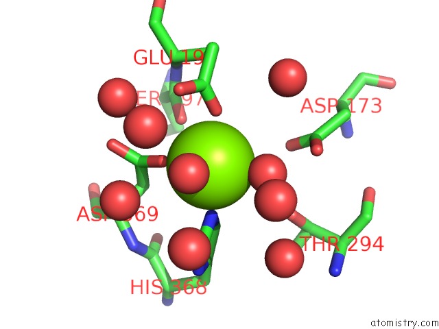
Mono view
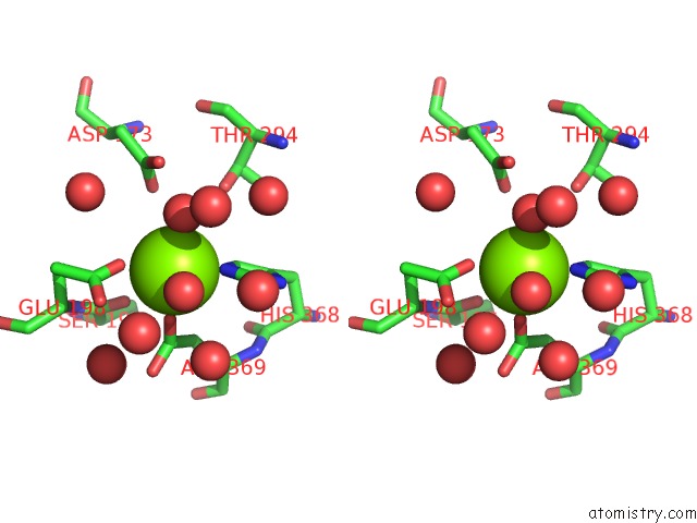
Stereo pair view
Mono view
Stereo pair view
A full contact list of Magnesium with other atoms in the Mg binding
site number 1 of X-Ray Structure of Cyanide-Bound Bovine Heart Cytochrome C Oxidase in the Fully Oxidized State at 2.0 Angstrom Resolution within 5.0Å range:
|
Magnesium binding site 2 out of 2 in 3x2q
Go back to
Magnesium binding site 2 out
of 2 in the X-Ray Structure of Cyanide-Bound Bovine Heart Cytochrome C Oxidase in the Fully Oxidized State at 2.0 Angstrom Resolution
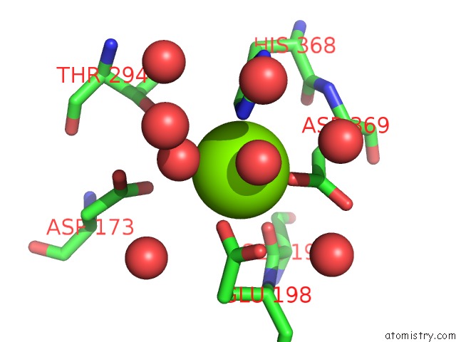
Mono view
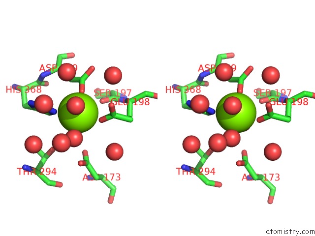
Stereo pair view
Mono view
Stereo pair view
A full contact list of Magnesium with other atoms in the Mg binding
site number 2 of X-Ray Structure of Cyanide-Bound Bovine Heart Cytochrome C Oxidase in the Fully Oxidized State at 2.0 Angstrom Resolution within 5.0Å range:
|
Reference:
N.Yano,
K.Muramoto,
M.Mochizuki,
K.Shinzawa-Itoh,
E.Yamashita,
S.Yoshikawa,
T.Tsukihara.
X-Ray Structure of Cyanide-Bound Bovine Heart Cytochrome C Oxidase in the Fully Oxidized State at 2.0 Angstrom Resolution. Acta Crystallogr F Struct V. 71 726 2015BIOL Commun.
ISSN: ESSN 2053-230X
PubMed: 26057802
DOI: 10.1107/S2053230X15007025
Page generated: Thu Aug 15 13:50:03 2024
ISSN: ESSN 2053-230X
PubMed: 26057802
DOI: 10.1107/S2053230X15007025
Last articles
Fe in 2YXOFe in 2YRS
Fe in 2YXC
Fe in 2YNM
Fe in 2YVJ
Fe in 2YP1
Fe in 2YU2
Fe in 2YU1
Fe in 2YQB
Fe in 2YOO