Magnesium »
PDB 3x1w-3zi8 »
3zi4 »
Magnesium in PDB 3zi4: The Structure of Beta-Phosphoglucomutase Inhibited with Glucose-6-Phospahte and Scandium Tetrafluoride
Enzymatic activity of The Structure of Beta-Phosphoglucomutase Inhibited with Glucose-6-Phospahte and Scandium Tetrafluoride
All present enzymatic activity of The Structure of Beta-Phosphoglucomutase Inhibited with Glucose-6-Phospahte and Scandium Tetrafluoride:
5.4.2.6;
5.4.2.6;
Protein crystallography data
The structure of The Structure of Beta-Phosphoglucomutase Inhibited with Glucose-6-Phospahte and Scandium Tetrafluoride, PDB code: 3zi4
was solved by
E.Pellegrini,
M.W.Bowler,
with X-Ray Crystallography technique. A brief refinement statistics is given in the table below:
| Resolution Low / High (Å) | 20.00 / 1.33 |
| Space group | P 21 21 21 |
| Cell size a, b, c (Å), α, β, γ (°) | 37.152, 54.238, 104.550, 90.00, 90.00, 90.00 |
| R / Rfree (%) | 13.018 / 17.379 |
Other elements in 3zi4:
The structure of The Structure of Beta-Phosphoglucomutase Inhibited with Glucose-6-Phospahte and Scandium Tetrafluoride also contains other interesting chemical elements:
| Scandium | (Sc) | 1 atom |
| Fluorine | (F) | 4 atoms |
Magnesium Binding Sites:
The binding sites of Magnesium atom in the The Structure of Beta-Phosphoglucomutase Inhibited with Glucose-6-Phospahte and Scandium Tetrafluoride
(pdb code 3zi4). This binding sites where shown within
5.0 Angstroms radius around Magnesium atom.
In total only one binding site of Magnesium was determined in the The Structure of Beta-Phosphoglucomutase Inhibited with Glucose-6-Phospahte and Scandium Tetrafluoride, PDB code: 3zi4:
In total only one binding site of Magnesium was determined in the The Structure of Beta-Phosphoglucomutase Inhibited with Glucose-6-Phospahte and Scandium Tetrafluoride, PDB code: 3zi4:
Magnesium binding site 1 out of 1 in 3zi4
Go back to
Magnesium binding site 1 out
of 1 in the The Structure of Beta-Phosphoglucomutase Inhibited with Glucose-6-Phospahte and Scandium Tetrafluoride
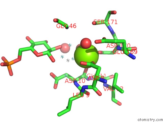
Mono view
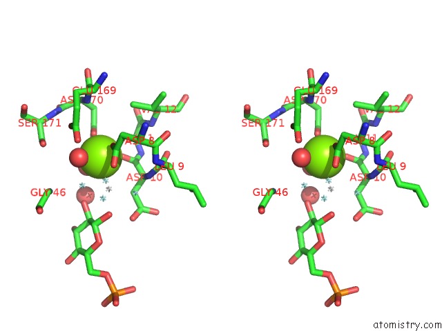
Stereo pair view
Mono view
Stereo pair view
A full contact list of Magnesium with other atoms in the Mg binding
site number 1 of The Structure of Beta-Phosphoglucomutase Inhibited with Glucose-6-Phospahte and Scandium Tetrafluoride within 5.0Å range:
|
Reference:
M.W.Bowler,
E.Pellegrini.
Metal Fluorides: Multi-Functional Tools For the Study of Phosphoryl Transfer Enzymes To Be Published.
Page generated: Thu Aug 15 13:55:31 2024
Last articles
F in 4JA8F in 4JBY
F in 4JB4
F in 4JAS
F in 4JA2
F in 4J3J
F in 4J8W
F in 4J8M
F in 4J6A
F in 4J6I