Magnesium »
PDB 3zx5-4a2b »
3zxd »
Magnesium in PDB 3zxd: Wild-Type Lysenin
Protein crystallography data
The structure of Wild-Type Lysenin, PDB code: 3zxd
was solved by
L.De Colibus,
A.F.P.Sonnen,
K.J.Morris,
C.A.Siebert,
P.Abrusci,
J.Plitzko,
V.Hodnik,
M.Leippe,
E.Volpi,
G.Anderluh,
R.J.C.Gilbert,
with X-Ray Crystallography technique. A brief refinement statistics is given in the table below:
| Resolution Low / High (Å) | 29.09 / 3.30 |
| Space group | P 1 |
| Cell size a, b, c (Å), α, β, γ (°) | 58.910, 85.560, 108.810, 98.88, 96.84, 90.04 |
| R / Rfree (%) | 21.3 / 23.6 |
Other elements in 3zxd:
The structure of Wild-Type Lysenin also contains other interesting chemical elements:
| Chlorine | (Cl) | 1 atom |
| Sodium | (Na) | 6 atoms |
Magnesium Binding Sites:
The binding sites of Magnesium atom in the Wild-Type Lysenin
(pdb code 3zxd). This binding sites where shown within
5.0 Angstroms radius around Magnesium atom.
In total 6 binding sites of Magnesium where determined in the Wild-Type Lysenin, PDB code: 3zxd:
Jump to Magnesium binding site number: 1; 2; 3; 4; 5; 6;
In total 6 binding sites of Magnesium where determined in the Wild-Type Lysenin, PDB code: 3zxd:
Jump to Magnesium binding site number: 1; 2; 3; 4; 5; 6;
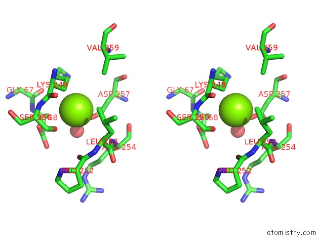
Magnesium binding site 1 out of 6 in 3zxd
Go back to
Magnesium binding site 1 out
of 6 in the Wild-Type Lysenin
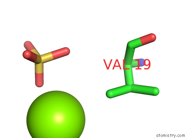
Mono view

Stereo pair view
Mono view
Stereo pair view
A full contact list of Magnesium with other atoms in the Mg binding
site number 1 of Wild-Type Lysenin within 5.0Å range:
|
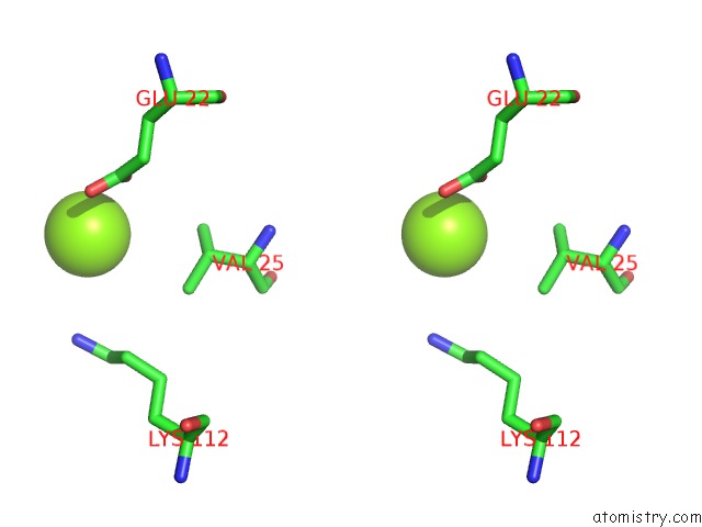
Magnesium binding site 2 out of 6 in 3zxd
Go back to
Magnesium binding site 2 out
of 6 in the Wild-Type Lysenin
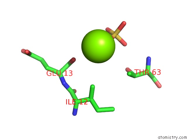
Mono view
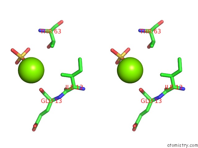
Stereo pair view
Mono view
Stereo pair view
A full contact list of Magnesium with other atoms in the Mg binding
site number 2 of Wild-Type Lysenin within 5.0Å range:
|
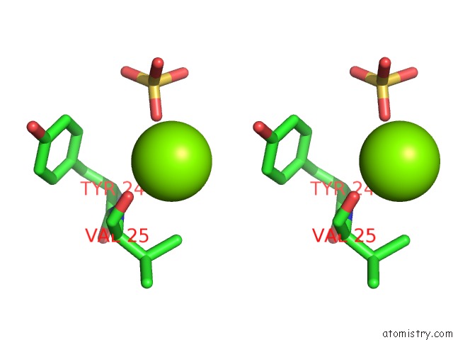
Magnesium binding site 3 out of 6 in 3zxd
Go back to
Magnesium binding site 3 out
of 6 in the Wild-Type Lysenin
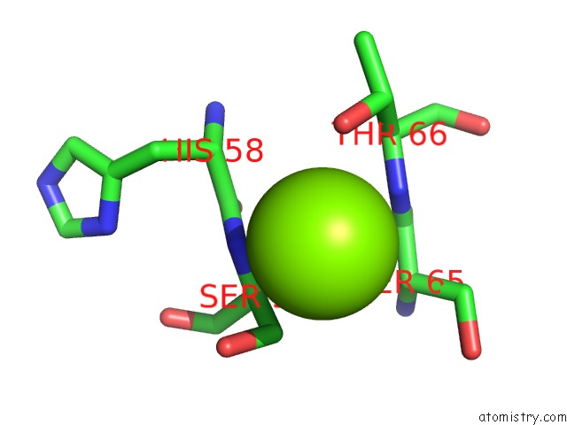
Mono view

Stereo pair view
Mono view
Stereo pair view
A full contact list of Magnesium with other atoms in the Mg binding
site number 3 of Wild-Type Lysenin within 5.0Å range:
|
Magnesium binding site 4 out of 6 in 3zxd
Go back to
Magnesium binding site 4 out
of 6 in the Wild-Type Lysenin

Mono view
Stereo pair view
Mono view
Stereo pair view
A full contact list of Magnesium with other atoms in the Mg binding
site number 4 of Wild-Type Lysenin within 5.0Å range:
|
Magnesium binding site 5 out of 6 in 3zxd
Go back to
Magnesium binding site 5 out
of 6 in the Wild-Type Lysenin

Mono view
Stereo pair view
Mono view
Stereo pair view
A full contact list of Magnesium with other atoms in the Mg binding
site number 5 of Wild-Type Lysenin within 5.0Å range:
|
Magnesium binding site 6 out of 6 in 3zxd
Go back to
Magnesium binding site 6 out
of 6 in the Wild-Type Lysenin

Mono view
Stereo pair view
Mono view
Stereo pair view
A full contact list of Magnesium with other atoms in the Mg binding
site number 6 of Wild-Type Lysenin within 5.0Å range:
|
Reference:
L.De Colibus,
A.F.P.Sonnen,
K.J.Morris,
C.A.Siebert,
P.Abrusci,
J.Plitzko,
V.Hodnik,
M.Leippe,
E.Volpi,
G.Anderluh,
R.J.C.Gilbert.
Structures of Lysenin Reveal A Shared Evolutionary Origin For Pore-Forming Proteins and Its Mode of Sphingomyelin Recognition. Structure V. 20 1498 2012.
ISSN: ISSN 0969-2126
PubMed: 22819216
DOI: 10.1016/J.STR.2012.06.011
Page generated: Thu Aug 15 14:08:57 2024
ISSN: ISSN 0969-2126
PubMed: 22819216
DOI: 10.1016/J.STR.2012.06.011
Last articles
Zn in 9MJ5Zn in 9HNW
Zn in 9G0L
Zn in 9FNE
Zn in 9DZN
Zn in 9E0I
Zn in 9D32
Zn in 9DAK
Zn in 8ZXC
Zn in 8ZUF