Magnesium »
PDB 3zx5-4a2b »
437d »
Magnesium in PDB 437d: Crystal Structure of An Rna Pseudoknot From Beet Western Yellow Virus Involved in Ribosomal Frameshifting
Protein crystallography data
The structure of Crystal Structure of An Rna Pseudoknot From Beet Western Yellow Virus Involved in Ribosomal Frameshifting, PDB code: 437d
was solved by
L.Su,
L.Chen,
M.Egli,
J.M.Berger,
A.Rich,
with X-Ray Crystallography technique. A brief refinement statistics is given in the table below:
| Resolution Low / High (Å) | 8.00 / 1.60 |
| Space group | P 32 2 1 |
| Cell size a, b, c (Å), α, β, γ (°) | 30.080, 30.080, 140.080, 90.00, 90.00, 120.00 |
| R / Rfree (%) | 20.7 / 25.4 |
Other elements in 437d:
The structure of Crystal Structure of An Rna Pseudoknot From Beet Western Yellow Virus Involved in Ribosomal Frameshifting also contains other interesting chemical elements:
| Sodium | (Na) | 1 atom |
Magnesium Binding Sites:
The binding sites of Magnesium atom in the Crystal Structure of An Rna Pseudoknot From Beet Western Yellow Virus Involved in Ribosomal Frameshifting
(pdb code 437d). This binding sites where shown within
5.0 Angstroms radius around Magnesium atom.
In total only one binding site of Magnesium was determined in the Crystal Structure of An Rna Pseudoknot From Beet Western Yellow Virus Involved in Ribosomal Frameshifting, PDB code: 437d:
In total only one binding site of Magnesium was determined in the Crystal Structure of An Rna Pseudoknot From Beet Western Yellow Virus Involved in Ribosomal Frameshifting, PDB code: 437d:
Magnesium binding site 1 out of 1 in 437d
Go back to
Magnesium binding site 1 out
of 1 in the Crystal Structure of An Rna Pseudoknot From Beet Western Yellow Virus Involved in Ribosomal Frameshifting
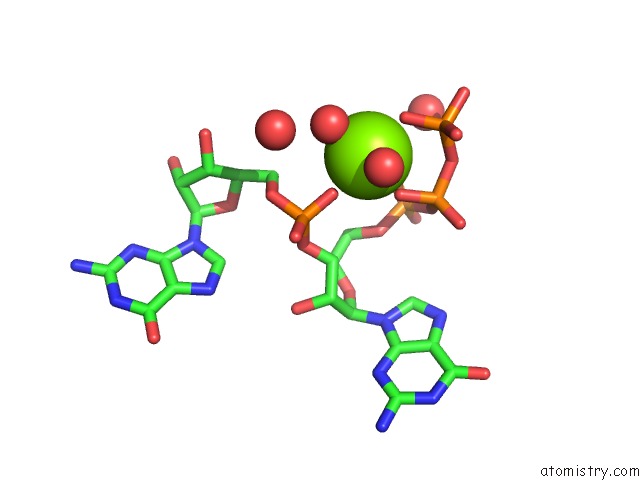
Mono view
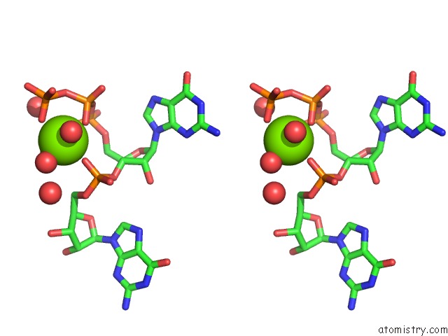
Stereo pair view
Mono view
Stereo pair view
A full contact list of Magnesium with other atoms in the Mg binding
site number 1 of Crystal Structure of An Rna Pseudoknot From Beet Western Yellow Virus Involved in Ribosomal Frameshifting within 5.0Å range:
|
Reference:
L.Su,
L.Chen,
M.Egli,
J.M.Berger,
A.Rich.
Minor Groove Rna Triplex in the Crystal Structure of A Ribosomal Frameshifting Viral Pseudoknot. Nat.Struct.Biol. V. 6 285 1999.
ISSN: ISSN 1072-8368
PubMed: 10074948
DOI: 10.1038/6722
Page generated: Thu Aug 15 14:13:10 2024
ISSN: ISSN 1072-8368
PubMed: 10074948
DOI: 10.1038/6722
Last articles
Fe in 2YXOFe in 2YRS
Fe in 2YXC
Fe in 2YNM
Fe in 2YVJ
Fe in 2YP1
Fe in 2YU2
Fe in 2YU1
Fe in 2YQB
Fe in 2YOO