Magnesium »
PDB 3zx5-4a2b »
462d »
Magnesium in PDB 462d: Crystal Structure of the Hiv-1 Genomic Rna Dimerization Initiation Site
Protein crystallography data
The structure of Crystal Structure of the Hiv-1 Genomic Rna Dimerization Initiation Site, PDB code: 462d
was solved by
E.Ennifar,
M.Yusupov,
P.Walter,
R.Marquet,
C.Ehresmann,
B.Ehresmann,
P.Dumas,
with X-Ray Crystallography technique. A brief refinement statistics is given in the table below:
| Resolution Low / High (Å) | 10.00 / 2.30 |
| Space group | P 31 2 1 |
| Cell size a, b, c (Å), α, β, γ (°) | 59.100, 59.100, 64.000, 90.00, 90.00, 120.00 |
| R / Rfree (%) | 21.1 / 21.2 |
Magnesium Binding Sites:
The binding sites of Magnesium atom in the Crystal Structure of the Hiv-1 Genomic Rna Dimerization Initiation Site
(pdb code 462d). This binding sites where shown within
5.0 Angstroms radius around Magnesium atom.
In total 8 binding sites of Magnesium where determined in the Crystal Structure of the Hiv-1 Genomic Rna Dimerization Initiation Site, PDB code: 462d:
Jump to Magnesium binding site number: 1; 2; 3; 4; 5; 6; 7; 8;
In total 8 binding sites of Magnesium where determined in the Crystal Structure of the Hiv-1 Genomic Rna Dimerization Initiation Site, PDB code: 462d:
Jump to Magnesium binding site number: 1; 2; 3; 4; 5; 6; 7; 8;
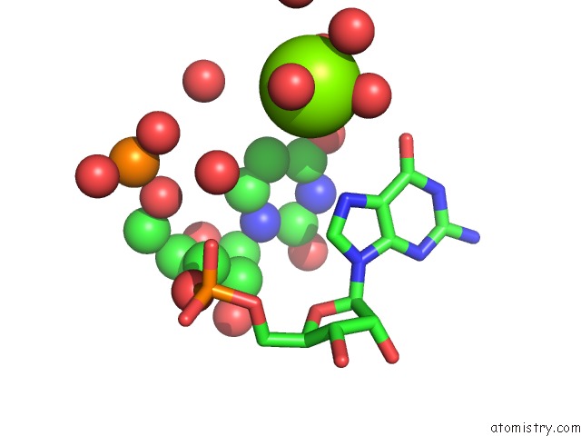
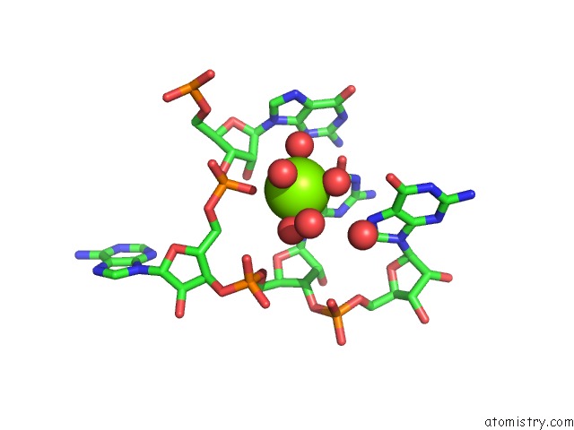
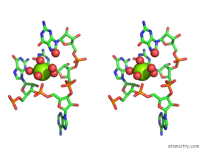
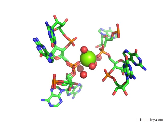
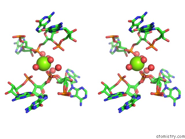
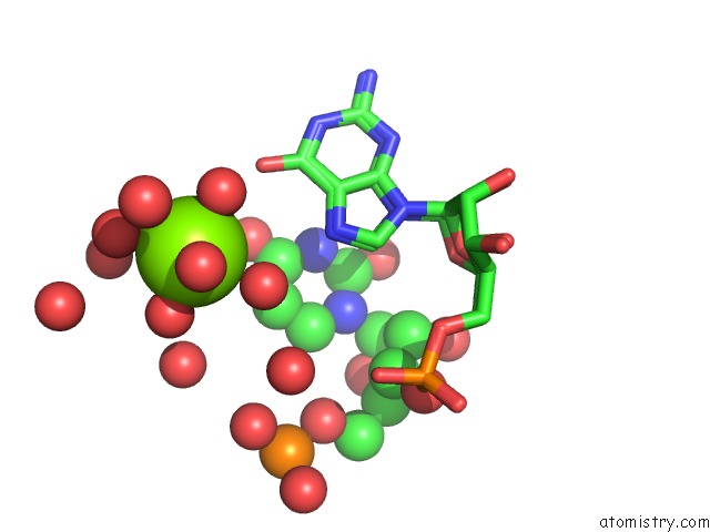
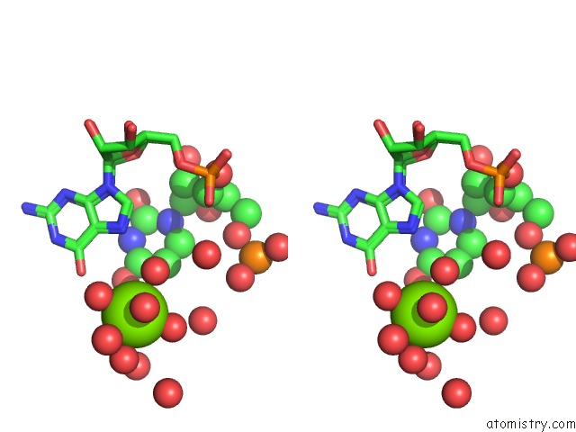
Magnesium binding site 1 out of 8 in 462d
Go back to
Magnesium binding site 1 out
of 8 in the Crystal Structure of the Hiv-1 Genomic Rna Dimerization Initiation Site
Mono view
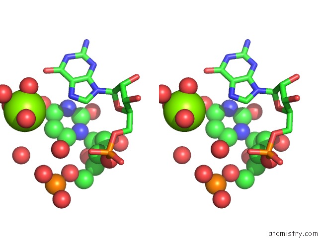
Stereo pair view
Mono view
Stereo pair view
A full contact list of Magnesium with other atoms in the Mg binding
site number 1 of Crystal Structure of the Hiv-1 Genomic Rna Dimerization Initiation Site within 5.0Å range:
|
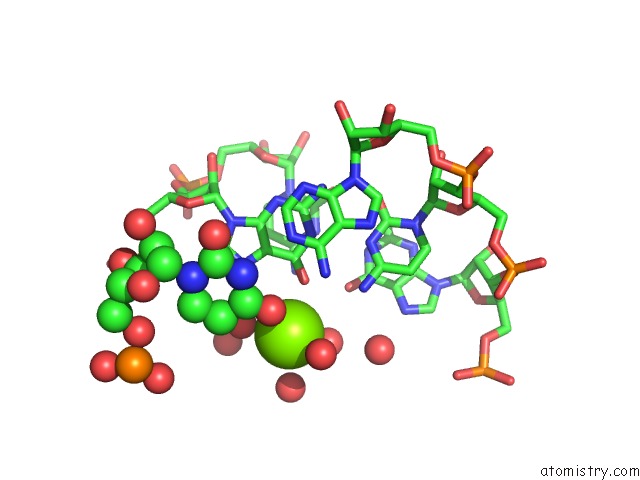
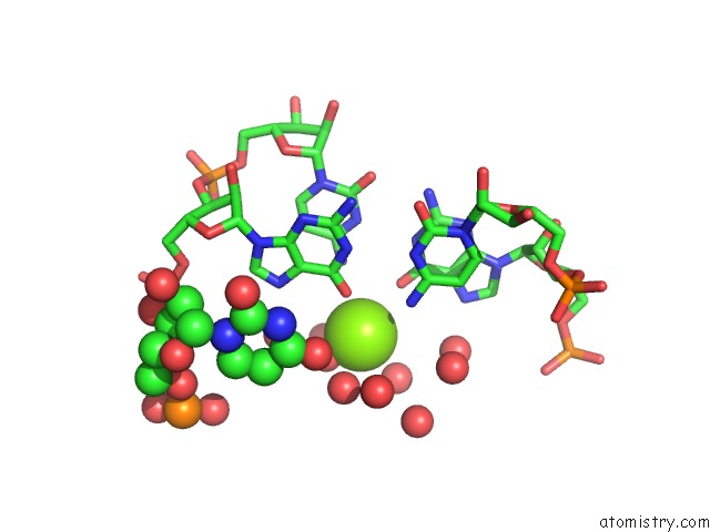
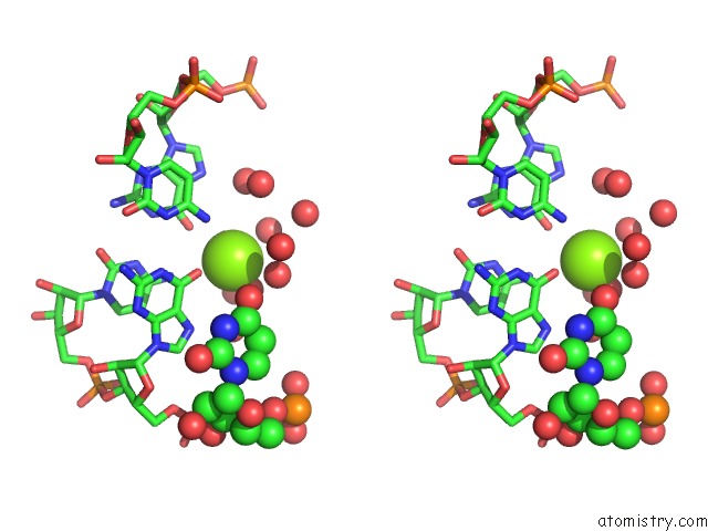
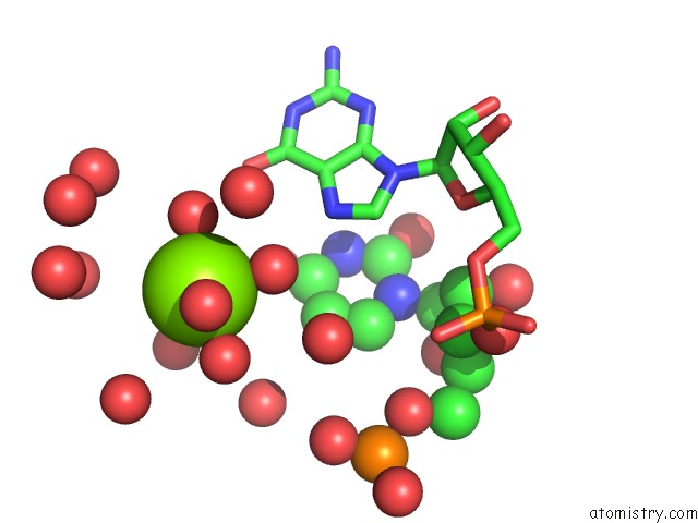
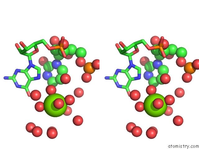
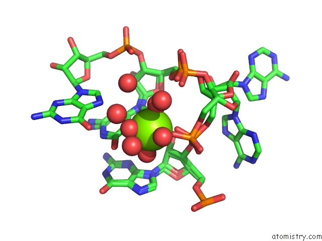
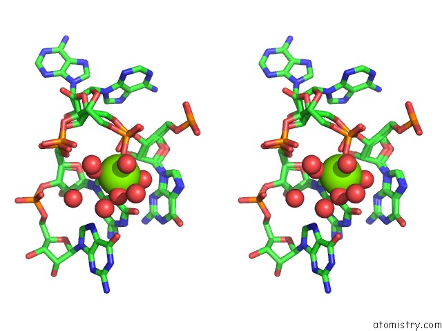
Magnesium binding site 2 out of 8 in 462d
Go back to
Magnesium binding site 2 out
of 8 in the Crystal Structure of the Hiv-1 Genomic Rna Dimerization Initiation Site
Mono view
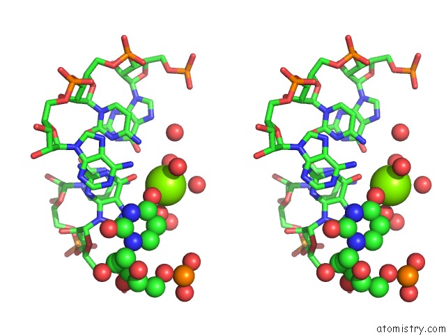
Stereo pair view
Mono view
Stereo pair view
A full contact list of Magnesium with other atoms in the Mg binding
site number 2 of Crystal Structure of the Hiv-1 Genomic Rna Dimerization Initiation Site within 5.0Å range:
|
Magnesium binding site 3 out of 8 in 462d
Go back to
Magnesium binding site 3 out
of 8 in the Crystal Structure of the Hiv-1 Genomic Rna Dimerization Initiation Site
Mono view
Stereo pair view
Mono view
Stereo pair view
A full contact list of Magnesium with other atoms in the Mg binding
site number 3 of Crystal Structure of the Hiv-1 Genomic Rna Dimerization Initiation Site within 5.0Å range:
|
Magnesium binding site 4 out of 8 in 462d
Go back to
Magnesium binding site 4 out
of 8 in the Crystal Structure of the Hiv-1 Genomic Rna Dimerization Initiation Site
Mono view
Stereo pair view
Mono view
Stereo pair view
A full contact list of Magnesium with other atoms in the Mg binding
site number 4 of Crystal Structure of the Hiv-1 Genomic Rna Dimerization Initiation Site within 5.0Å range:
|
Magnesium binding site 5 out of 8 in 462d
Go back to
Magnesium binding site 5 out
of 8 in the Crystal Structure of the Hiv-1 Genomic Rna Dimerization Initiation Site
Mono view
Stereo pair view
Mono view
Stereo pair view
A full contact list of Magnesium with other atoms in the Mg binding
site number 5 of Crystal Structure of the Hiv-1 Genomic Rna Dimerization Initiation Site within 5.0Å range:
|
Magnesium binding site 6 out of 8 in 462d
Go back to
Magnesium binding site 6 out
of 8 in the Crystal Structure of the Hiv-1 Genomic Rna Dimerization Initiation Site
Mono view
Stereo pair view
Mono view
Stereo pair view
A full contact list of Magnesium with other atoms in the Mg binding
site number 6 of Crystal Structure of the Hiv-1 Genomic Rna Dimerization Initiation Site within 5.0Å range:
|
Magnesium binding site 7 out of 8 in 462d
Go back to
Magnesium binding site 7 out
of 8 in the Crystal Structure of the Hiv-1 Genomic Rna Dimerization Initiation Site
Mono view
Stereo pair view
Mono view
Stereo pair view
A full contact list of Magnesium with other atoms in the Mg binding
site number 7 of Crystal Structure of the Hiv-1 Genomic Rna Dimerization Initiation Site within 5.0Å range:
|
Magnesium binding site 8 out of 8 in 462d
Go back to
Magnesium binding site 8 out
of 8 in the Crystal Structure of the Hiv-1 Genomic Rna Dimerization Initiation Site
Mono view
Stereo pair view
Mono view
Stereo pair view
A full contact list of Magnesium with other atoms in the Mg binding
site number 8 of Crystal Structure of the Hiv-1 Genomic Rna Dimerization Initiation Site within 5.0Å range:
|
Reference:
E.Ennifar,
M.Yusupov,
P.Walter,
R.Marquet,
B.Ehresmann,
C.Ehresmann,
P.Dumas.
The Crystal Structure of the Dimerization Initiation Site of Genomic Hiv-1 Rna Reveals An Extended Duplex with Two Adenine Bulges. Structure Fold.Des. V. 7 1439 1999.
ISSN: ISSN 0969-2126
PubMed: 10574792
DOI: 10.1016/S0969-2126(00)80033-7
Page generated: Mon Aug 11 05:29:35 2025
ISSN: ISSN 0969-2126
PubMed: 10574792
DOI: 10.1016/S0969-2126(00)80033-7
Last articles
Mg in 4FNJMg in 4FPP
Mg in 4FP1
Mg in 4FO6
Mg in 4FO0
Mg in 4FME
Mg in 4FMM
Mg in 4FNI
Mg in 4FMO
Mg in 4FMD