Magnesium »
PDB 4a2b-4aar »
4a50 »
Magnesium in PDB 4a50: Crystal Structure of Human Kinesin EG5 in Complex with 2-Amino-5-(3-Methylphenyl)-5,5-Diphenylpentanoic Acid
Protein crystallography data
The structure of Crystal Structure of Human Kinesin EG5 in Complex with 2-Amino-5-(3-Methylphenyl)-5,5-Diphenylpentanoic Acid, PDB code: 4a50
was solved by
H.Y.K.Kaan,
F.Kozielski,
with X-Ray Crystallography technique. A brief refinement statistics is given in the table below:
| Resolution Low / High (Å) | 28.03 / 2.75 |
| Space group | I 21 3 |
| Cell size a, b, c (Å), α, β, γ (°) | 158.540, 158.540, 158.540, 90.00, 90.00, 90.00 |
| R / Rfree (%) | 19.759 / 24.301 |
Magnesium Binding Sites:
The binding sites of Magnesium atom in the Crystal Structure of Human Kinesin EG5 in Complex with 2-Amino-5-(3-Methylphenyl)-5,5-Diphenylpentanoic Acid
(pdb code 4a50). This binding sites where shown within
5.0 Angstroms radius around Magnesium atom.
In total only one binding site of Magnesium was determined in the Crystal Structure of Human Kinesin EG5 in Complex with 2-Amino-5-(3-Methylphenyl)-5,5-Diphenylpentanoic Acid, PDB code: 4a50:
In total only one binding site of Magnesium was determined in the Crystal Structure of Human Kinesin EG5 in Complex with 2-Amino-5-(3-Methylphenyl)-5,5-Diphenylpentanoic Acid, PDB code: 4a50:
Magnesium binding site 1 out of 1 in 4a50
Go back to
Magnesium binding site 1 out
of 1 in the Crystal Structure of Human Kinesin EG5 in Complex with 2-Amino-5-(3-Methylphenyl)-5,5-Diphenylpentanoic Acid
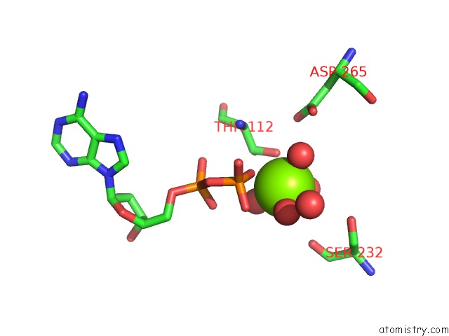
Mono view
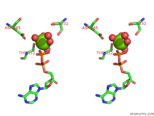
Stereo pair view
Mono view
Stereo pair view
A full contact list of Magnesium with other atoms in the Mg binding
site number 1 of Crystal Structure of Human Kinesin EG5 in Complex with 2-Amino-5-(3-Methylphenyl)-5,5-Diphenylpentanoic Acid within 5.0Å range:
|
Reference:
F.Wang,
J.A.D.Good,
O.Rath,
H.Y.K.Kaan,
O.B.Sutcliffe,
S.P.Mackay,
F.Kozielski.
Triphenylbutanamines: Kinesin Spindle Protein Inhibitors with in Vivo Antitumor Activity. J.Med.Chem. V. 55 1511 2012.
ISSN: ISSN 0022-2623
PubMed: 22248262
DOI: 10.1021/JM201195M
Page generated: Thu Aug 15 14:20:37 2024
ISSN: ISSN 0022-2623
PubMed: 22248262
DOI: 10.1021/JM201195M
Last articles
Zn in 9JYWZn in 9IR4
Zn in 9IR3
Zn in 9GMX
Zn in 9GMW
Zn in 9JEJ
Zn in 9ERF
Zn in 9ERE
Zn in 9EGV
Zn in 9EGW